• 文献检索
  • 文档翻译
  • 深度研究
  • 学术资讯
  • Suppr Zotero 插件Zotero 插件
  • 邀请有礼
  • 套餐&价格
  • 历史记录
应用&插件
Suppr Zotero 插件Zotero 插件浏览器插件Mac 客户端Windows 客户端微信小程序
定价
高级版会员购买积分包购买API积分包
服务
文献检索文档翻译深度研究API 文档MCP 服务
关于我们
关于 Suppr公司介绍联系我们用户协议隐私条款
关注我们

Suppr 超能文献

核心技术专利:CN118964589B侵权必究
粤ICP备2023148730 号-1Suppr @ 2026

文献检索

告别复杂PubMed语法,用中文像聊天一样搜索,搜遍4000万医学文献。AI智能推荐,让科研检索更轻松。

立即免费搜索

文件翻译

保留排版,准确专业,支持PDF/Word/PPT等文件格式,支持 12+语言互译。

免费翻译文档

深度研究

AI帮你快速写综述,25分钟生成高质量综述,智能提取关键信息,辅助科研写作。

立即免费体验

偏振傅里叶变换红外光谱揭示细菌视紫红质中功能性水分子的定位与取向

Localization and orientation of functional water molecules in bacteriorhodopsin as revealed by polarized Fourier transform infrared spectroscopy.

作者信息

Hatanaka M, Kandori H, Maeda A

机构信息

Department of Biophysics, Graduate School of Science, Kyoto University, Japan.

出版信息

Biophys J. 1997 Aug;73(2):1001-6. doi: 10.1016/S0006-3495(97)78133-5.

DOI:10.1016/S0006-3495(97)78133-5
PMID:9251817
原文链接:https://pmc.ncbi.nlm.nih.gov/articles/PMC1180997/
Abstract

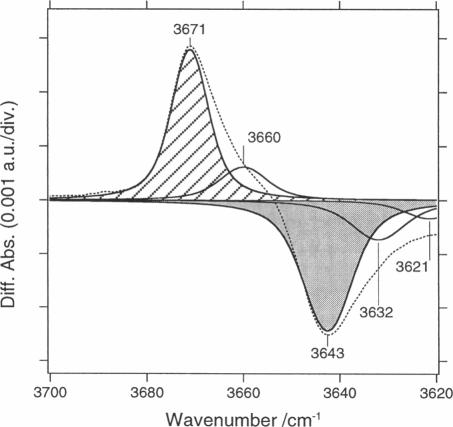
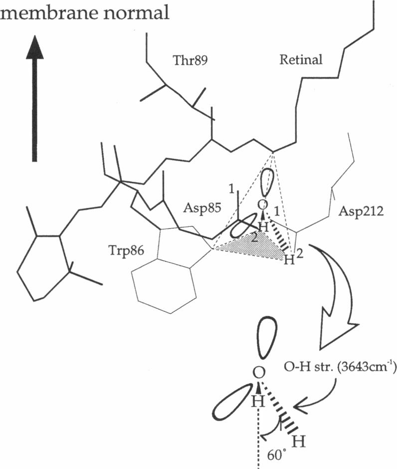
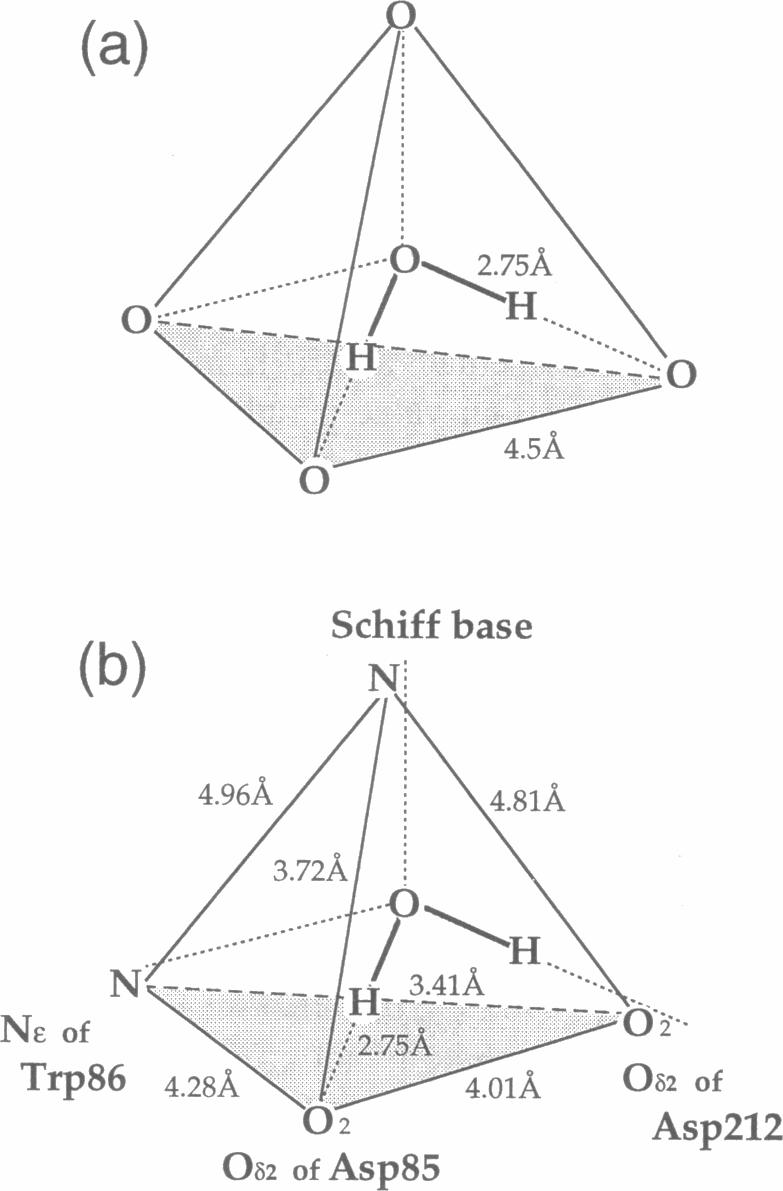
Linear dichroic difference Fourier transform infrared spectra upon formation of the M photointermediate were recorded with oriented purple membranes. The purpose was to determine the angle of the directions of the dipole moments of 1) the water molecule whose O-H stretching vibration appears at 3643 cm-1 for the unphotolyzed state and 3671 cm-1 for the M intermediate, and 2) the C=O bond of protonated Asp85 in the M intermediate. The angle of 36 degrees we find for the C=O of the protonated Asp85 in the M intermediate is not markedly different from 26 degrees for unprotonated Asp85 in the model based on cryoelectron diffraction, indicating the absence of gross orientation changes in Asp85 upon its protonation. The O-H band at 3671 cm-1 of a water molecule in the M intermediate, although its position has not determined, is fixed almost parallel to the membrane plane. For the unphotolyzed state the angle of the water O-H to the membrane normal was determined to be 60 degrees. On the basis of these data and the structural model, we place the water molecule in the unphotolyzed state at a position where it forms hydrogen bonds with the Schiff base, Asp85, Asp212, and Trp86.

摘要

用取向紫膜记录了形成M光中间体时的线性二色性差示傅里叶变换红外光谱。目的是确定以下两种情况偶极矩方向的夹角:1)未光解状态下O-H伸缩振动出现在3643cm-1处、M中间体中出现在3671cm-1处的水分子;2)M中间体中质子化的Asp85的C=O键。我们发现M中间体中质子化的Asp85的C=O键夹角为36度,这与基于低温电子衍射的模型中未质子化的Asp85的26度夹角没有明显差异,表明Asp85质子化时没有明显的取向变化。M中间体中一个水分子在3671cm-1处的O-H带,尽管其位置尚未确定,但几乎与膜平面平行。对于未光解状态,水分子O-H与膜法线的夹角确定为60度。基于这些数据和结构模型,我们将未光解状态下的水分子置于与席夫碱、Asp85、Asp212和Trp86形成氢键的位置。

https://cdn.ncbi.nlm.nih.gov/pmc/blobs/7dd4/1180997/19db0ba5aab4/biophysj00033-0457-a.jpg
https://cdn.ncbi.nlm.nih.gov/pmc/blobs/7dd4/1180997/d74565c39361/biophysj00033-0455-a.jpg
https://cdn.ncbi.nlm.nih.gov/pmc/blobs/7dd4/1180997/30c291680eb6/biophysj00033-0456-a.jpg
https://cdn.ncbi.nlm.nih.gov/pmc/blobs/7dd4/1180997/19db0ba5aab4/biophysj00033-0457-a.jpg
https://cdn.ncbi.nlm.nih.gov/pmc/blobs/7dd4/1180997/d74565c39361/biophysj00033-0455-a.jpg
https://cdn.ncbi.nlm.nih.gov/pmc/blobs/7dd4/1180997/30c291680eb6/biophysj00033-0456-a.jpg
https://cdn.ncbi.nlm.nih.gov/pmc/blobs/7dd4/1180997/19db0ba5aab4/biophysj00033-0457-a.jpg

相似文献

1
Localization and orientation of functional water molecules in bacteriorhodopsin as revealed by polarized Fourier transform infrared spectroscopy.偏振傅里叶变换红外光谱揭示细菌视紫红质中功能性水分子的定位与取向
Biophys J. 1997 Aug;73(2):1001-6. doi: 10.1016/S0006-3495(97)78133-5.
2
Water molecules in the schiff base region of bacteriorhodopsin.细菌视紫红质席夫碱区域中的水分子。
J Am Chem Soc. 2003 Nov 5;125(44):13312-3. doi: 10.1021/ja037343s.
3
Hydration switch model for the proton transfer in the Schiff base region of bacteriorhodopsin.细菌视紫红质席夫碱区域质子转移的水合开关模型
Biochim Biophys Acta. 2004 Jul 23;1658(1-2):72-9. doi: 10.1016/j.bbabio.2004.03.015.
4
Hydration of the counterion of the Schiff base in the chloride-transporting mutant of bacteriorhodopsin: FTIR and FT-raman studies of the effects of anion binding when Asp85 is replaced with a neutral residue.细菌视紫红质氯离子转运突变体中席夫碱抗衡离子的水合作用:当天冬氨酸85被中性残基取代时阴离子结合效应的傅里叶变换红外光谱和傅里叶变换拉曼光谱研究
Biochemistry. 1996 Nov 12;35(45):14244-50. doi: 10.1021/bi9606197.
5
Structural changes of water in the Schiff base region of bacteriorhodopsin: proposal of a hydration switch model.细菌视紫红质席夫碱区域水的结构变化:水合开关模型的提出
Biochemistry. 2003 Mar 4;42(8):2300-6. doi: 10.1021/bi026990d.
6
Hydrogen bonds of water and C==O groups coordinate long-range structural changes in the L photointermediate of bacteriorhodopsin.水的氢键和C==O基团协调细菌视紫红质L光中间体中的长程结构变化。
Biochemistry. 1996 Apr 2;35(13):4063-8. doi: 10.1021/bi9524530.
7
Interaction of aspartate-85 with a water molecule and the protonated Schiff base in the L intermediate of bacteriorhodopsin: a Fourier-transform infrared spectroscopic study.细菌视紫红质L中间体中天门冬氨酸85与水分子及质子化席夫碱的相互作用:傅里叶变换红外光谱研究
Biochemistry. 1994 Feb 22;33(7):1713-7. doi: 10.1021/bi00173a013.
8
Water structural changes in the L and M photocycle intermediates of bacteriorhodopsin as revealed by time-resolved step-scan Fourier transform infrared (FTIR) spectroscopy.时间分辨步进扫描傅里叶变换红外(FTIR)光谱揭示的细菌视紫红质L和M光循环中间体中的水结构变化。
Biochemistry. 2007 Mar 13;46(10):2787-96. doi: 10.1021/bi0616596. Epub 2007 Feb 15.
9
FTIR studies of internal water molecules in the Schiff base region of bacteriorhodopsin.细菌视紫红质席夫碱区域内水分子的傅里叶变换红外光谱研究。
Biochemistry. 2005 May 24;44(20):7406-13. doi: 10.1021/bi050122+.
10
Water-mediated hydrogen-bonded network on the cytoplasmic side of the Schiff base of the L photointermediate of bacteriorhodopsin.细菌视紫红质L光中间体席夫碱细胞质侧的水介导氢键网络。
Biochemistry. 2003 Dec 9;42(48):14122-9. doi: 10.1021/bi0301542.

引用本文的文献

1
Allosteric Effects of the Proton Donor on the Microbial Proton Pump Proteorhodopsin.质子供体对微生物质子泵蛋白视紫红质的变构效应。
Biophys J. 2018 Oct 2;115(7):1240-1250. doi: 10.1016/j.bpj.2018.08.028. Epub 2018 Aug 29.
2
Conformational changes in the archaerhodopsin-3 proton pump: detection of conserved strongly hydrogen bonded water networks.古紫质-3质子泵的构象变化:保守的强氢键水网络的检测
J Biol Phys. 2012 Jan;38(1):153-68. doi: 10.1007/s10867-011-9246-4. Epub 2011 Dec 10.
3
Factors influencing the energetics of electron and proton transfers in proteins. What can be learned from calculations.

本文引用的文献

1
Redshift of the purple membrane absorption band and the deprotonation of tyrosine residues at high pH: Origin of the parallel photocycles of trans-bacteriorhodopsin.高 pH 值下紫膜吸收带的红移和酪氨酸残基的去质子化:反细菌视紫红质平行光循环的起源。
Biophys J. 1991 Aug;60(2):475-90. doi: 10.1016/S0006-3495(91)82074-4.
2
Trp86 --> Phe replacement in bacteriorhodopsin affects a water molecule near Asp85 and light adaptation.
Biochemistry. 1997 May 6;36(18):5493-8. doi: 10.1021/bi970081k.
3
Intramembrane signaling mediated by hydrogen-bonding of water and carboxyl groups in bacteriorhodopsin and rhodopsin.
J Biochem. 1997 Mar;121(3):399-406. doi: 10.1093/oxfordjournals.jbchem.a021602.
影响蛋白质中电子和质子转移能量学的因素。从计算中能学到什么。
Biochim Biophys Acta. 2006 Aug;1757(8):942-68. doi: 10.1016/j.bbabio.2006.06.005. Epub 2006 Jun 17.
4
Structural changes in bacteriorhodopsin during the photocycle measured by time-resolved polarized Fourier transform infrared spectroscopy.通过时间分辨偏振傅里叶变换红外光谱法测量细菌视紫红质在光循环过程中的结构变化。
Biophys J. 2001 Dec;81(6):3577-89. doi: 10.1016/S0006-3495(01)75988-7.
5
Tight Asp-85--Thr-89 association during the pump switch of bacteriorhodopsin.细菌视紫红质泵转换过程中紧密的天冬氨酸85-苏氨酸89关联
Proc Natl Acad Sci U S A. 2001 Feb 13;98(4):1571-6. doi: 10.1073/pnas.98.4.1571.
6
Local and distant protein structural changes on photoisomerization of the retinal in bacteriorhodopsin.细菌视紫红质中视黄醛光异构化时的局部和远距离蛋白质结构变化。
Proc Natl Acad Sci U S A. 2000 Apr 25;97(9):4643-8. doi: 10.1073/pnas.080064797.
7
Azide reduces the hydrophobic barrier of the bacteriorhodopsin proton channel.叠氮化物降低了细菌视紫红质子通道的疏水屏障。
Biophys J. 1999 May;76(5):2702-10. doi: 10.1016/S0006-3495(99)77422-9.
4
Hydration of the counterion of the Schiff base in the chloride-transporting mutant of bacteriorhodopsin: FTIR and FT-raman studies of the effects of anion binding when Asp85 is replaced with a neutral residue.细菌视紫红质氯离子转运突变体中席夫碱抗衡离子的水合作用:当天冬氨酸85被中性残基取代时阴离子结合效应的傅里叶变换红外光谱和傅里叶变换拉曼光谱研究
Biochemistry. 1996 Nov 12;35(45):14244-50. doi: 10.1021/bi9606197.
5
Thermodynamic stability of water molecules in the bacteriorhodopsin proton channel: a molecular dynamics free energy perturbation study.细菌视紫红质质子通道中水分子的热力学稳定性:分子动力学自由能微扰研究
Biophys J. 1996 Aug;71(2):670-81. doi: 10.1016/S0006-3495(96)79267-6.
6
Electron-crystallographic refinement of the structure of bacteriorhodopsin.细菌视紫红质结构的电子晶体学精修
J Mol Biol. 1996 Jun 14;259(3):393-421. doi: 10.1006/jmbi.1996.0328.
7
Effects of arginine-82 on the interactions of internal water molecules in bacteriorhodopsin.精氨酸-82对细菌视紫红质中内部水分子相互作用的影响。
Biochemistry. 1996 May 21;35(20):6308-12. doi: 10.1021/bi952973v.
8
Molecular dynamics study of the M412 intermediate of bacteriorhodopsin.细菌视紫红质M412中间体的分子动力学研究
Biophys J. 1995 Dec;69(6):2745-60. doi: 10.1016/S0006-3495(95)80146-3.
9
Molecular dynamics study of the proton pump cycle of bacteriorhodopsin.细菌视紫红质质子泵循环的分子动力学研究
Biochemistry. 1993 Mar 9;32(9):2291-306. doi: 10.1021/bi00060a022.
10
Proton translocation mechanism and energetics in the light-driven pump bacteriorhodopsin.光驱动泵视紫红质中的质子转运机制与能量学
Biochim Biophys Acta. 1993 Dec 7;1183(2):241-61. doi: 10.1016/0005-2728(93)90226-6.