Lawton D M, Andrew J G, Marsh D R, Hoyland J A, Freemont A J
Department of Pathological Sciences, University of Manchester, UK.
Mol Pathol. 1997 Aug;50(4):194-7. doi: 10.1136/mp.50.4.194.
High levels of collagen type III are biochemically detectable in biopsies of non-uniting fractures, and in the serum of patients suffering from this condition. The aim of this study was to determine whether the expression of collagen type III was limited to fibrous tissue in non-unions, or whether some was present in bone.
Biopsies from normally healing human fractures and non-unions were examined using in situ hybridisation and immunohistochemistry.
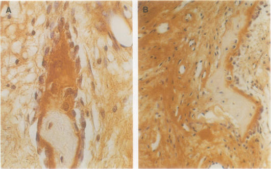
The mesenchymal cell population, which includes fibroblast and osteoblast precursors, expressed mRNA for collagen type III. However, mature osteoblasts on the surface of woven bone varied profoundly between normally healing fractures (in which they were negative or occasionally weakly positive) and non-unions (in which they were strongly positive). Areas of woven bone that had osteoblasts positive for collagen type III mRNA also immunostained positively for the protein.
This study shows that non-union fracture callus osteoblasts on the surfaces of woven bone exhibit an unusual phenotype: they express collagen type III, a molecule characteristic of an earlier stage of osteoblast differentiation, which is not expressed by osteoblasts on woven bone surfaces of bone that develops normally. This finding may be useful in developing an early clinical test for impending non-union.
在不愈合骨折的活检组织以及患有该病症患者的血清中,可通过生化检测到高水平的III型胶原蛋白。本研究的目的是确定III型胶原蛋白的表达是否仅限于不愈合部位的纤维组织,还是在骨组织中也有存在。
使用原位杂交和免疫组织化学方法检查正常愈合的人类骨折和不愈合骨折的活检组织。
包括成纤维细胞和成骨细胞前体的间充质细胞群体表达III型胶原蛋白的mRNA。然而,编织骨表面的成熟成骨细胞在正常愈合骨折(其中它们为阴性或偶尔弱阳性)和不愈合骨折(其中它们为强阳性)之间有很大差异。III型胶原蛋白mRNA呈阳性的成骨细胞所在的编织骨区域,其蛋白质免疫染色也呈阳性。
本研究表明,编织骨表面的不愈合骨折骨痂成骨细胞表现出一种不寻常的表型:它们表达III型胶原蛋白,这是成骨细胞分化早期阶段的一种特征性分子,而在正常发育的骨组织编织骨表面的成骨细胞中不表达。这一发现可能有助于开发针对即将发生的不愈合的早期临床试验。