Olveczky B P, Periasamy N, Verkman A S
Department of Medicine, Cardiovascular Research Institute, University of California, San Francisco 94143, USA.
Biophys J. 1997 Nov;73(5):2836-47. doi: 10.1016/S0006-3495(97)78312-7.
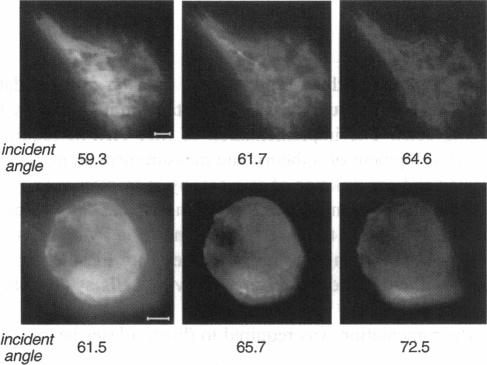
The decay of evanescent field intensity beyond a dielectric interface depends upon beam incident angle, enabling the 3-d distribution of fluorophores to be deduced from total internal reflection fluorescence microscopy (TIRFM) images obtained at multiple incident angles. Instrumentation was constructed for computer-automated multiple angle-TIRFM (MA-TIRFM) using a right angle F2 glass prism (n(r) 1.632) to create the dielectric interface. A laser beam (488 nm) was attenuated by an acoustooptic modulator and directed onto a specified spot on the prism surface. Beam incident angle was set using three microstepper motors controlling two rotatable mirrors and a rotatable optical flat. TIRFM images were acquired by a cooled CCD camera in approximately 0.5 degree steps for >15 incident angles starting from the critical angle. For cell studies, cells were grown directly on the glass prisms (without refractive index-matching fluid) and positioned in the optical path. Images of the samples were acquired at multiple angles, and corrected for angle-dependent evanescent field intensity using "reference" images acquired with a fluorophore solution replacing the sample. A theory was developed to compute fluorophore z-distribution by inverse Laplace transform of angle-resolved intensity functions. The theory included analysis of multiple layers of different refractive index for cell studies, and the anisotropic emission from fluorophores near a dielectric interface. Instrument performance was validated by mapping the thickness of a film of dihexyloxacarbocyanine in DMSO/water (n(r) 1.463) between the F2 glass prism and a plano-convex silica lens (458 mm radius, n(r) 1.463); the MA-TIRFM map accurately reproduced the lens spherical surface. MA-TIRFM was used to compare with nanometer z-resolution the geometry of cell-substrate contact for BCECF-labeled 3T3 fibroblasts versus MDCK epithelial cells. These studies establish MA-TIRFM for measurement of submicroscopic distances between fluorescent probes and cell membranes.
超越介电界面的倏逝场强度衰减取决于光束入射角,这使得能够从在多个入射角获得的全内反射荧光显微镜(TIRFM)图像中推断出荧光团的三维分布。使用直角F2玻璃棱镜(n(r) 1.632)构建了用于计算机自动化多角度TIRFM(MA-TIRFM)的仪器,以创建介电界面。激光束(488 nm)由声光调制器衰减并导向棱镜表面上的指定点。使用控制两个可旋转镜和一个可旋转光学平面的三个微步电机设置光束入射角。从临界角开始,由冷却的CCD相机以约0.5度的步长采集>15个入射角的TIRFM图像。对于细胞研究,细胞直接生长在玻璃棱镜上(不使用折射率匹配液)并置于光路中。在多个角度采集样品图像,并使用用荧光团溶液代替样品采集的“参考”图像校正角度相关的倏逝场强度。通过对角度分辨强度函数进行拉普拉斯逆变换,开发了一种计算荧光团z分布的理论。该理论包括对用于细胞研究的不同折射率多层的分析,以及介电界面附近荧光团的各向异性发射。通过绘制F2玻璃棱镜和平凸透镜(半径458 mm,n(r) 1.463)之间二己基氧杂碳菁在DMSO/水中(n(r) 1.463)薄膜的厚度来验证仪器性能;MA-TIRFM图准确再现了透镜球面。使用MA-TIRFM将BCECF标记的3T3成纤维细胞与MDCK上皮细胞的细胞-基质接触几何结构与纳米z分辨率进行比较。这些研究确立了MA-TIRFM用于测量荧光探针与细胞膜之间的亚微观距离。