Frolov I, Houri-Hadad Y, Soskolne A, Shapira L
Department of Periodontology, Hebrew University-Hadassah School of Dental Medicine, Jerusalem, Israel.
Immunology. 1998 Mar;93(3):323-8. doi: 10.1046/j.1365-2567.1998.00437.x.
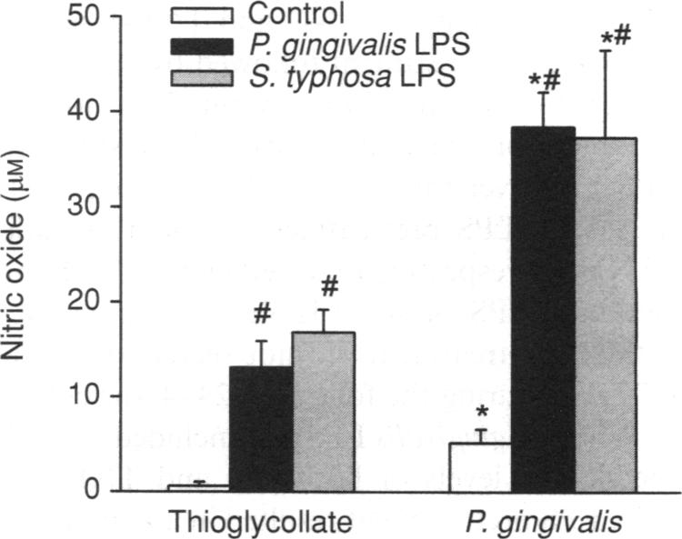
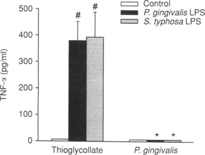
The present study was designed to test whether the functional response of mouse macrophages elicited by chronic exposure to bacteria will be different from that of cells elicited by a non-bacterial irritant. Macrophage elicitation was conducted by Porphyromonas gingivalis, a major periodontal pathogen, in comparison to a standard elicitation by thioglycollate (TG). We measured lipopolysaccharide (LPS)-induced nitric oxide (NO) and tumour necrosis factor-alpha (TNF-alpha) secretion by the elicited macrophages, and the expression of inflammatory cytokines in the whole elicited cell population. In addition, we tested the response of TG-elicited macrophages to pretreatment with P. gingivalis LPS in vitro. Mouse peritoneal macrophages were harvested 4 days after intraperitoneal injection of TG or heat-killed P. gingivalis. TG-elicited macrophages produced undetectable levels of TNF-alpha and approximately 0.5 microM of NO. The stimulation of the macrophages with LPS resulted in the secretion of NO and TNF-alpha in a dose-dependent manner. The P. gingivalis-elicited macrophages produced basal levels of approximately 5 microM NO, but TNF-alpha was not detectable. LPS stimulation of these cells further increased the secretion of NO eightfold while TNF-alpha remained undetectable. The NO secretion by P. gingivalis-elicited cells was significantly higher than that by TG-elicited cells. Examination of cytokine expression in the whole elicited cell population revealed that both P. gingivalis-elicited cells and TG-elicited cells expressed messenger RNA for interleukin-2 (IL-2), TNF-alpha and interferon-gamma (IFN-gamma), but not for IL-4. IL-6 was expressed in P. gingivalis-elicited cells only. Pretreatment of TG-elicited macrophages with P. gingivalis LPS for 24 hr prior to a second LPS challenge resulted in down-regulation of TNF-alpha secretion and up-regulation of NO secretion, a response similar to that seen in P. gingivalis-elicited peritoneal macrophages. The results suggest that the in vivo exposure of resident macrophages to P. gingivalis induces functional changes in peritoneal macrophages. These changes might be due to the effect of P. gingivalis LPS.
本研究旨在测试长期暴露于细菌所引发的小鼠巨噬细胞的功能反应是否会与由非细菌性刺激物引发的细胞的功能反应有所不同。与用巯基乙酸盐(TG)进行的标准刺激相比,巨噬细胞的诱导是通过主要的牙周病原体牙龈卟啉单胞菌来进行的。我们测量了诱导的巨噬细胞中脂多糖(LPS)诱导的一氧化氮(NO)和肿瘤坏死因子-α(TNF-α)的分泌,以及整个诱导细胞群体中炎性细胞因子的表达。此外,我们在体外测试了TG诱导的巨噬细胞对牙龈卟啉单胞菌LPS预处理的反应。在腹腔注射TG或热灭活的牙龈卟啉单胞菌4天后收获小鼠腹腔巨噬细胞。TG诱导的巨噬细胞产生的TNF-α水平检测不到,产生的NO约为0.5微摩尔。用LPS刺激巨噬细胞会导致NO和TNF-α以剂量依赖性方式分泌。牙龈卟啉单胞菌诱导的巨噬细胞产生的基础水平的NO约为5微摩尔,但检测不到TNF-α。用LPS刺激这些细胞会使NO的分泌进一步增加八倍,而TNF-α仍然检测不到。牙龈卟啉单胞菌诱导的细胞分泌的NO明显高于TG诱导的细胞。对整个诱导细胞群体中的细胞因子表达进行检测发现,牙龈卟啉单胞菌诱导的细胞和TG诱导的细胞都表达白细胞介素-2(IL-2)、TNF-α和干扰素-γ(IFN-γ)的信使RNA,但不表达IL-4的信使RNA。IL-6仅在牙龈卟啉单胞菌诱导的细胞中表达。在第二次LPS刺激前24小时用牙龈卟啉单胞菌LPS对TG诱导的巨噬细胞进行预处理,会导致TNF-α分泌下调和NO分泌上调,这一反应与牙龈卟啉单胞菌诱导的腹腔巨噬细胞中观察到的反应相似。结果表明,驻留巨噬细胞在体内暴露于牙龈卟啉单胞菌会诱导腹腔巨噬细胞发生功能变化。这些变化可能是由于牙龈卟啉单胞菌LPS的作用。