Virág L, Scott G S, Cuzzocrea S, Marmer D, Salzman A L, Szabó C
Division of Critical Care, Children's Hospital Medical Center, Cincinnati, OH 45229, USA.
Immunology. 1998 Jul;94(3):345-55. doi: 10.1046/j.1365-2567.1998.00534.x.
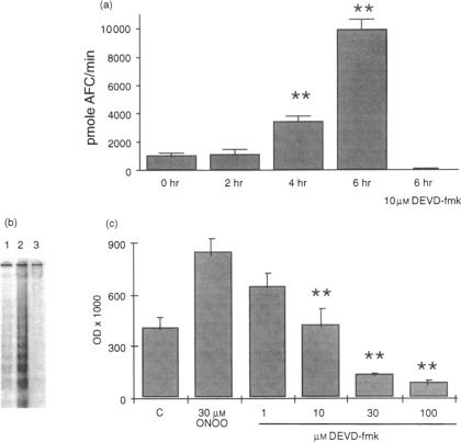
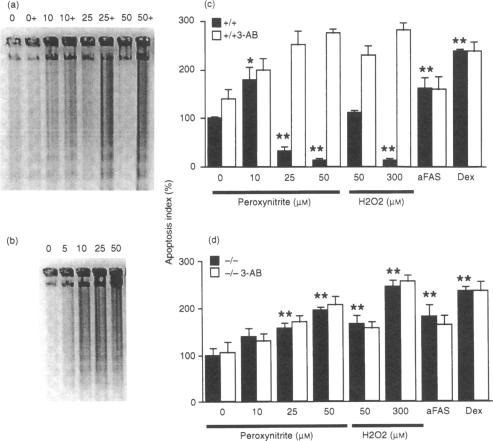
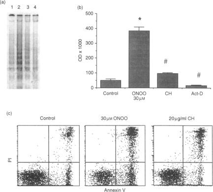
The mechanisms by which immature thymocyte apoptosis is induced during negative selection are poorly defined. Reports demonstrated that cross-linking of T-cell receptor leads to stromal cell activation, expression of inducible nitric oxide synthase (iNOS) and, subsequently, to thymocyte apoptosis. Therefore we examined, whether NO directly or indirectly, through peroxynitrite formation, causes thymocyte apoptosis. Immuno-histochemical detection of nitrotyrosine revealed in vivo peroxynitrite formation in the thymi of naive mice. Nitrotyrosine, the footprint of peroxynitrite, was predominantly found in the corticomedullary junction and the medulla of naive mice. In the thymi of mice deficient in the inducible isoform of nitric oxide synthase, considerably less nitrotyrosine was found. Exposure of thymocytes in vitro to low concentrations (10 microM) of peroxynitrite led to apoptosis, whereas higher concentrations (50 microM) resulted in intense cell death with the characteristics of necrosis. We also investigated the effect of poly (ADP-ribose) synthetase (PARS) inhibition on thymocyte apoptosis. Using the PARS inhibitor 3-aminobenzamide (3-AB), or thymocytes from PARS-deficient animals, we established that PARS determines the fate of thymocyte death. Suppression of cellular ATP levels, and the cellular necrosis in response to peroxynitrite were prevented by PARS inhibition. Therefore, in the absence of PARS, cells are diverted towards the pathway of apoptotic cell death. Similar results were obtained with H2O2 treatment, while apoptosis induced by non-oxidative stimuli such as dexamethasone or anti-FAS antibody was unaffected by PARS inhibition. In conclusion, we propose that peroxynitrite-induced apoptosis may play a role in the process of thymocyte negative selection. Furthermore, we propose that the physiological role of PARS cleavage by apopain during apoptosis may serve as an energy-conserving step, enabling the cell to complete the process of apoptosis.
在阴性选择过程中诱导未成熟胸腺细胞凋亡的机制仍不清楚。报告表明,T细胞受体的交联导致基质细胞活化、诱导型一氧化氮合酶(iNOS)的表达,随后导致胸腺细胞凋亡。因此,我们研究了一氧化氮是否直接或通过过氧亚硝酸盐的形成间接导致胸腺细胞凋亡。免疫组织化学检测硝基酪氨酸显示,在未接触过抗原的小鼠胸腺中存在过氧亚硝酸盐的体内形成。硝基酪氨酸,即过氧亚硝酸盐的痕迹,主要存在于未接触过抗原的小鼠的皮质髓质交界处和髓质中。在缺乏诱导型一氧化氮合酶同工型的小鼠胸腺中,发现的硝基酪氨酸明显较少。体外将胸腺细胞暴露于低浓度(10微摩尔)的过氧亚硝酸盐会导致凋亡,而较高浓度(50微摩尔)则会导致具有坏死特征的强烈细胞死亡。我们还研究了聚(ADP-核糖)合成酶(PARS)抑制对胸腺细胞凋亡的影响。使用PARS抑制剂3-氨基苯甲酰胺(3-AB)或来自PARS缺陷动物的胸腺细胞,我们确定PARS决定胸腺细胞死亡的命运。PARS抑制可防止细胞ATP水平的降低以及对过氧亚硝酸盐的细胞坏死反应。因此,在没有PARS的情况下,细胞会转向凋亡性细胞死亡途径。用H2O2处理也得到了类似的结果,而由地塞米松或抗FAS抗体等非氧化刺激诱导的凋亡不受PARS抑制的影响。总之,我们认为过氧亚硝酸盐诱导的凋亡可能在胸腺细胞阴性选择过程中起作用。此外,我们认为凋亡过程中凋亡蛋白酶对PARS的切割的生理作用可能是一个节能步骤,使细胞能够完成凋亡过程。