Shryock T R, Losonsky J M, Smith W C, Gatlin C L, Francisco C J, Kuriashkin I V, Clarkson R B, Jordan W H
Elanco Animal Health, Eli Lilly and Company, Greenfield, Indiana 46140, USA.
Can J Vet Res. 1998 Oct;62(4):287-92.
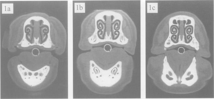
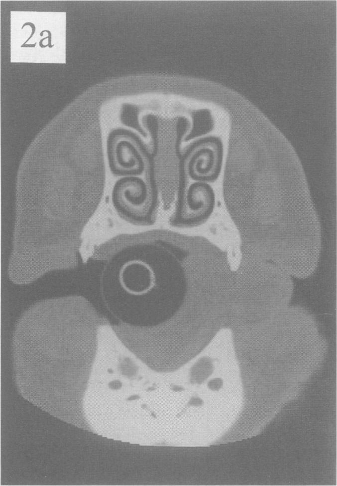
A non-invasive imaging modality, computed tomography (CT), was used to visualize changes in nasal turbinates of anesthetized pigs over a 12-week observation period (pigs were 14 wk of age at study week 0). Normal, non-infected pigs were compared to pigs with mild challenge-induced atrophic rhinitis (AR) in order to detect subtle differences in morphology. To determine feasibility for time course studies in future experiments, morphometric quantitation at the level of the 2nd premolar (turbinate area ratio or TAR) in cross-section CT images at multiple timepoints was done. Additionally, at study termination, the TAR determined from CT images, magnetic resonance imaging (MRI), and wet tissue (WT), were compared to each other and to the standard subjective measure, visual scoring. There were no statistically significant differences between the control and AR groups at CT imaging dates of 0, 3, 6, 9, or 12 wk (P = 0.182). However, a statistically significant decrease in TAR measurements over time (P = 0.015) was observed in both groups, with lower mean values observed on Weeks 3 and 6 before rebounding to baseline values at study termination. At Week 12 (termination of the study), the TAR measurements derived from CT, MRI, and WT were not statistically different from one another (P = 0.220) and the treatment group-by-method interaction was not significant (P = 0.800). This provided evidence of equivalency of the techniques. Mean values for normal and infected groups were not significantly different based on either TAR imaging methods (P = 0.552) or visual scores (P = 0.088). Thus, the current study demonstrated that CT was an acceptable alternative imaging modality which could be used for quantitation of turbinate changes in snouts of live pigs to provide data comparable to tissue taken at necropsy. Computed tomographic imaging would allow non-invasive tracking of disease or treatment responses within individual animals over time. Morphometric analysis of the TAR was equivalent between the CT, MRI, and WT specimens.
一种非侵入性成像方式——计算机断层扫描(CT),被用于在12周的观察期内观察麻醉猪鼻甲的变化(研究第0周时猪为14周龄)。将正常、未感染的猪与轻度激发性萎缩性鼻炎(AR)猪进行比较,以检测形态学上的细微差异。为了确定在未来实验中进行时间进程研究的可行性,在多个时间点对横断面CT图像中第二前磨牙水平的形态计量学定量(鼻甲面积比或TAR)进行了测定。此外,在研究结束时,将CT图像、磁共振成像(MRI)和湿组织(WT)测定的TAR相互比较,并与标准主观测量方法视觉评分进行比较。在CT成像日期为0、3、6、9或12周时,对照组和AR组之间无统计学显著差异(P = 0.182)。然而,两组均观察到TAR测量值随时间有统计学显著下降(P = 0.015),在第3周和第6周观察到较低的平均值,然后在研究结束时回升至基线值。在第12周(研究结束时),CT、MRI和WT得出的TAR测量值彼此之间无统计学差异(P = 0.220),且治疗组×方法的交互作用不显著(P = 0.800)。这提供了这些技术等效性的证据。基于TAR成像方法(P = 0.552)或视觉评分(P = 0.088),正常组和感染组的平均值无显著差异。因此,当前研究表明CT是一种可接受的替代成像方式,可用于定量活体猪口鼻部鼻甲变化,以提供与尸检时获取的组织相当的数据。计算机断层扫描成像将允许随时间对个体动物内的疾病或治疗反应进行非侵入性跟踪。CT、MRI和WT标本之间TAR的形态计量学分析是等效的。