• 文献检索
  • 文档翻译
  • 深度研究
  • 学术资讯
  • Suppr Zotero 插件Zotero 插件
  • 邀请有礼
  • 套餐&价格
  • 历史记录
应用&插件
Suppr Zotero 插件Zotero 插件浏览器插件Mac 客户端Windows 客户端微信小程序
定价
高级版会员购买积分包购买API积分包
服务
文献检索文档翻译深度研究API 文档MCP 服务
关于我们
关于 Suppr公司介绍联系我们用户协议隐私条款
关注我们

Suppr 超能文献

核心技术专利:CN118964589B侵权必究
粤ICP备2023148730 号-1Suppr @ 2026

文献检索

告别复杂PubMed语法,用中文像聊天一样搜索,搜遍4000万医学文献。AI智能推荐,让科研检索更轻松。

立即免费搜索

文件翻译

保留排版,准确专业,支持PDF/Word/PPT等文件格式,支持 12+语言互译。

免费翻译文档

深度研究

AI帮你快速写综述,25分钟生成高质量综述,智能提取关键信息,辅助科研写作。

立即免费体验

在人红白血病细胞系中,通过反义技术敲除肌醇1,3,4,5 - 四磷酸受体GAP1(IP4BP)会导致出现中间电导的钾钙通道,这些通道使细胞膜超极化并增强钙内流。

Antisense knock out of the inositol 1,3,4,5-tetrakisphosphate receptor GAP1(IP4BP) in the human erythroleukemia cell line leads to the appearance of intermediate conductance K(Ca) channels that hyperpolarize the membrane and enhance calcium influx.

作者信息

Lu X, Fein A, Feinstein M B, O'Rourke F A

机构信息

Department of Pharmacology, The University of Connecticut Health Center, Farmington, Connecticut 06030, USA.

出版信息

J Gen Physiol. 1999 Jan;113(1):81-96. doi: 10.1085/jgp.113.1.81.

DOI:10.1085/jgp.113.1.81
PMID:9874690
原文链接:https://pmc.ncbi.nlm.nih.gov/articles/PMC2222987/
Abstract

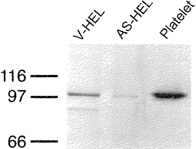
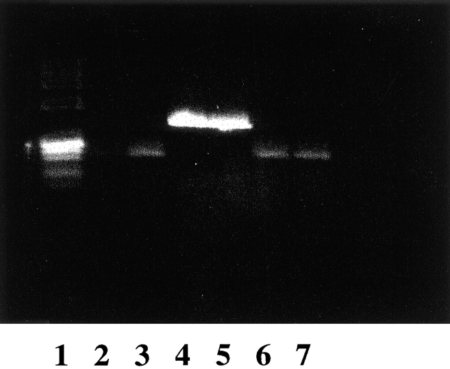
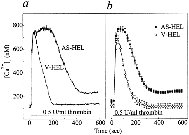
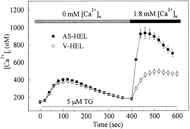
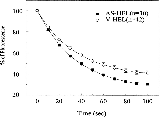
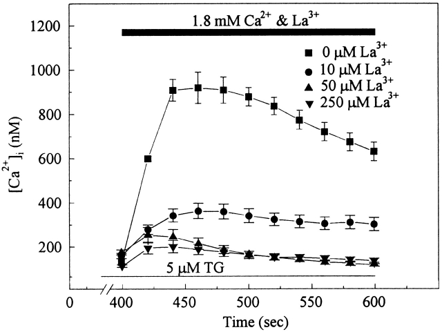
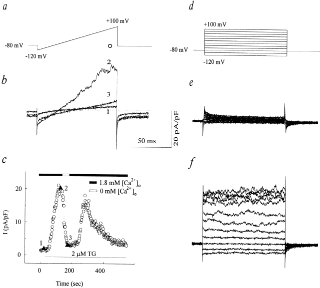
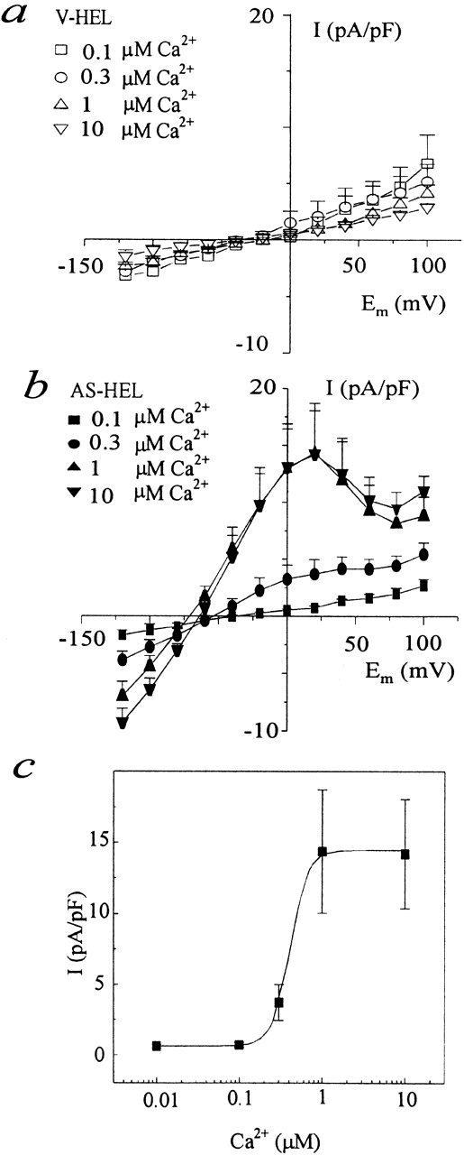
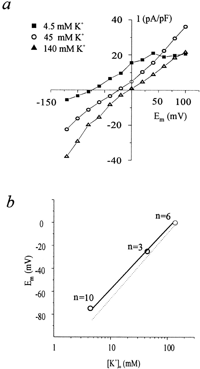
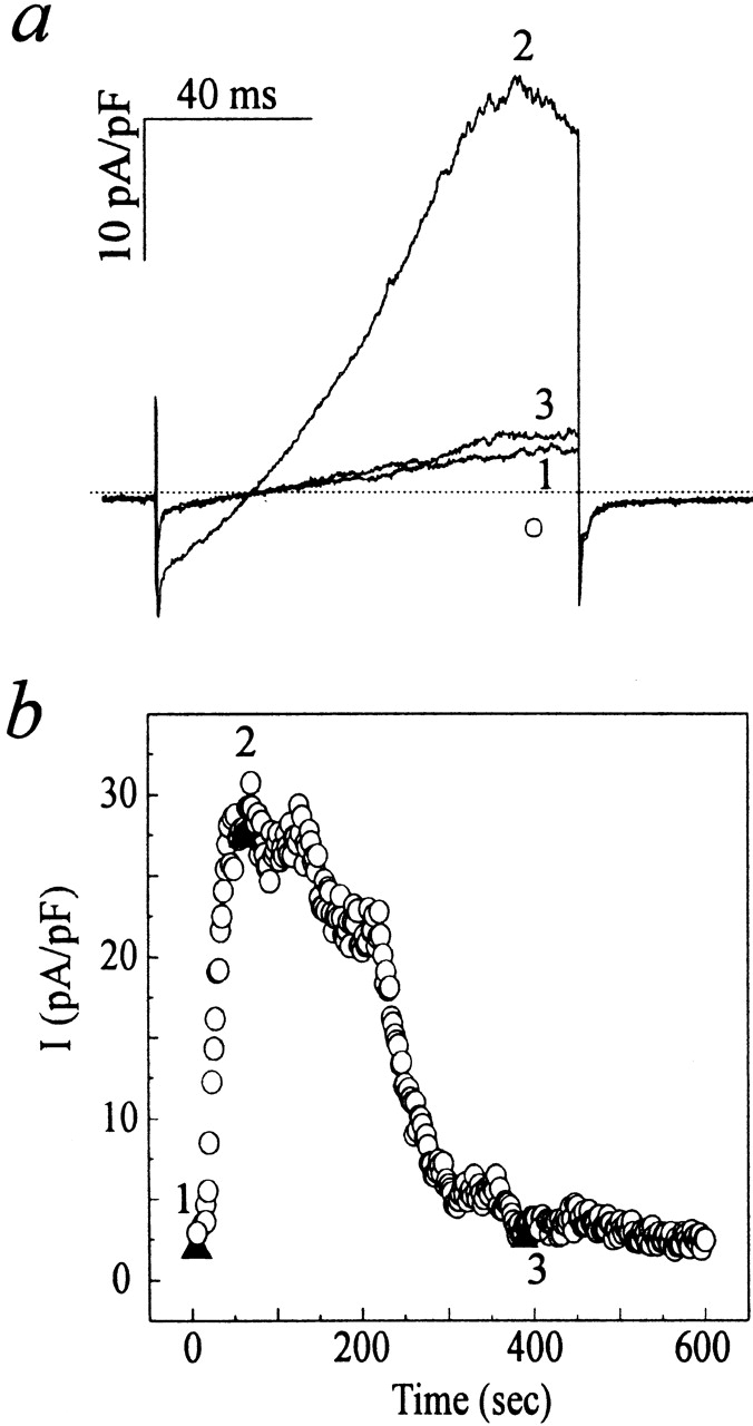
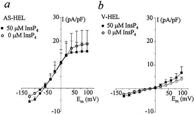
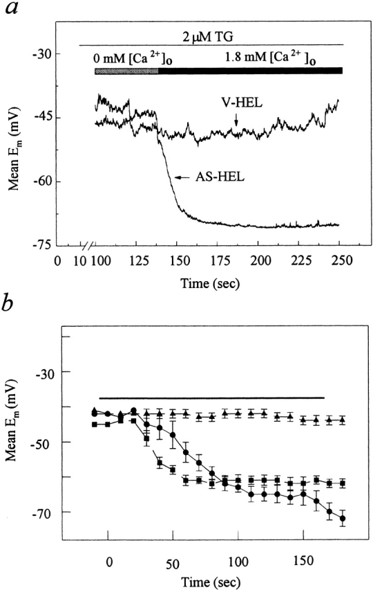
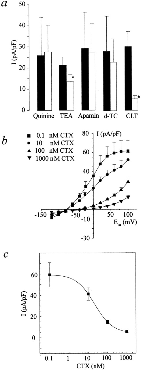
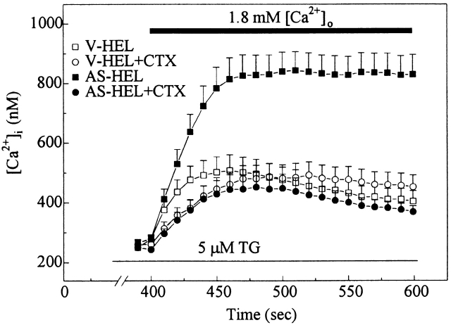
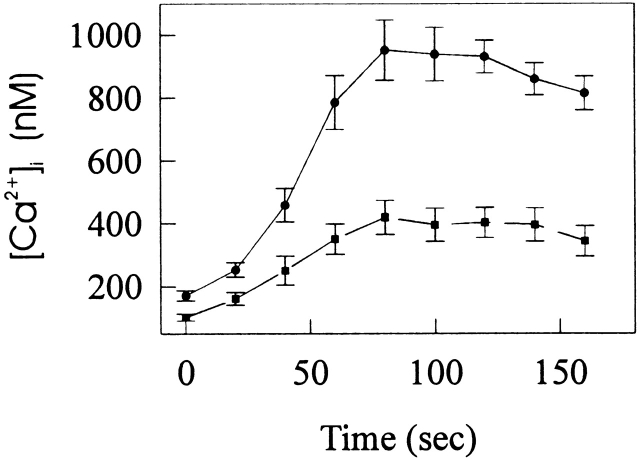
To study the role of the inositol 1,3,4,5-trisphosphate-binding protein GAP1(IP4BP) in store-operated Ca2+ entry, we established a human erythroleukemia (HEL) cell line in which the expression of GAP1(IP4BP) was substantially reduced by transfection with a vector containing antisense DNA under control of a Rous Sarcoma virus promoter and the Escherichia coli LacI repressor (AS-HEL cells). Control cells were transfected with vector lacking antisense DNA (V-HEL cells). GAP1(IP4BP) protein, which is a member of the GTPase-activating protein (GAP1) family, was reduced by 85% in AS-HEL cells and was further reduced by 96% by treatment with isopropylthio-beta-D- galactoside to relieve LacI repression. The loss of GAP1(IP4BP) was associated with both a membrane hyperpolarization and a substantially increased Ca2+ entry induced by thrombin or thapsigargin. The activation of intermediate conductance Ca2+-activated K+ channels in AS-HEL cells (not seen in V-HEL cells) was responsible for the membrane hyperpolarization and the enhanced Ca2+ entry, and both were blocked by charybdotoxin. Stimulated V-HEL cells did not hyperpolarize and basal Ca2+ influx was unaffected by charybdotoxin. In V-HEL cells hyperpolarized by removal of extracellular K+, the thapsigargin-stimulated Ca2+ influx was increased. Expression of mRNA for the human Ca2+-activated intermediate conductance channel KCa4 was equivalent in both AS-HEL and V-HEL cells, suggesting that the specific appearance of calcium-activated potassium current (IK(Ca)) in AS-HEL cells was possibly due to modulation of preexisting channels. Our results demonstrate that GAP1(IP4BP), likely working through a signaling pathway dependent on a small GTP-binding protein, can regulate the function of K(Ca) channels that produce a hyperpolarizing current that substantially enhances the magnitude and time course of Ca2+ entry subsequent to the release of internal Ca2+ stores.

摘要

为研究肌醇1,3,4,5 - 三磷酸结合蛋白GAP1(IP4BP)在储存性钙内流中的作用,我们建立了一种人红白血病(HEL)细胞系,该细胞系通过用含有在劳氏肉瘤病毒启动子和大肠杆菌LacI阻遏物控制下的反义DNA的载体转染,使GAP1(IP4BP)的表达大幅降低(AS - HEL细胞)。对照细胞用缺乏反义DNA的载体转染(V - HEL细胞)。GAP1(IP4BP)蛋白是GTP酶激活蛋白(GAP1)家族的成员,在AS - HEL细胞中减少了85%,并用异丙基硫代 - β - D - 半乳糖苷处理以解除LacI阻遏后进一步减少了96%。GAP1(IP4BP)的缺失与膜超极化以及凝血酶或毒胡萝卜素诱导的钙内流大幅增加有关。AS - HEL细胞中中等电导钙激活钾通道的激活(在V - HEL细胞中未观察到)导致了膜超极化和增强的钙内流,两者均被蝎毒素阻断。刺激的V - HEL细胞未发生超极化,基础钙内流不受蝎毒素影响。在通过去除细胞外钾而超极化的V - HEL细胞中,毒胡萝卜素刺激的钙内流增加。人钙激活中等电导通道KCa4的mRNA表达在AS - HEL细胞和V - HEL细胞中相当,这表明AS - HEL细胞中钙激活钾电流(IK(Ca))的特异性出现可能是由于对现有通道的调节。我们的结果表明,GAP1(IP4BP)可能通过依赖小GTP结合蛋白的信号通路发挥作用,可调节产生超极化电流的K(Ca)通道的功能,该电流在内部钙储存释放后可大幅增强钙内流的幅度和时间进程。

https://cdn.ncbi.nlm.nih.gov/pmc/blobs/9ca5/2222987/bcb66574a9e4/JGP7786.f15.jpg
https://cdn.ncbi.nlm.nih.gov/pmc/blobs/9ca5/2222987/1084b4d3641d/JGP7786.f1.jpg
https://cdn.ncbi.nlm.nih.gov/pmc/blobs/9ca5/2222987/2001d58fe394/JGP7786.f2.jpg
https://cdn.ncbi.nlm.nih.gov/pmc/blobs/9ca5/2222987/354bb87669ee/JGP7786.f3.jpg
https://cdn.ncbi.nlm.nih.gov/pmc/blobs/9ca5/2222987/b0dae61f739f/JGP7786.f4.jpg
https://cdn.ncbi.nlm.nih.gov/pmc/blobs/9ca5/2222987/e612ba582311/JGP7786.f5.jpg
https://cdn.ncbi.nlm.nih.gov/pmc/blobs/9ca5/2222987/c27e26dc94aa/JGP7786.f6.jpg
https://cdn.ncbi.nlm.nih.gov/pmc/blobs/9ca5/2222987/7226d99dffaa/JGP7786.f7.jpg
https://cdn.ncbi.nlm.nih.gov/pmc/blobs/9ca5/2222987/63180e685fc1/JGP7786.f8.jpg
https://cdn.ncbi.nlm.nih.gov/pmc/blobs/9ca5/2222987/dd670ee0f977/JGP7786.f9.jpg
https://cdn.ncbi.nlm.nih.gov/pmc/blobs/9ca5/2222987/9ab4471a3dbf/JGP7786.f10.jpg
https://cdn.ncbi.nlm.nih.gov/pmc/blobs/9ca5/2222987/7daf03aa1919/JGP7786.f11.jpg
https://cdn.ncbi.nlm.nih.gov/pmc/blobs/9ca5/2222987/5dede99233d7/JGP7786.f12.jpg
https://cdn.ncbi.nlm.nih.gov/pmc/blobs/9ca5/2222987/6b615b69a210/JGP7786.f13.jpg
https://cdn.ncbi.nlm.nih.gov/pmc/blobs/9ca5/2222987/555a38d30b85/JGP7786.f14.jpg
https://cdn.ncbi.nlm.nih.gov/pmc/blobs/9ca5/2222987/bcb66574a9e4/JGP7786.f15.jpg

相似文献

1
Antisense knock out of the inositol 1,3,4,5-tetrakisphosphate receptor GAP1(IP4BP) in the human erythroleukemia cell line leads to the appearance of intermediate conductance K(Ca) channels that hyperpolarize the membrane and enhance calcium influx.在人红白血病细胞系中,通过反义技术敲除肌醇1,3,4,5 - 四磷酸受体GAP1(IP4BP)会导致出现中间电导的钾钙通道,这些通道使细胞膜超极化并增强钙内流。
J Gen Physiol. 1999 Jan;113(1):81-96. doi: 10.1085/jgp.113.1.81.
2
Thrombin-dependent calcium signalling in single human erythroleukaemia cells.单个人类红白血病细胞中凝血酶依赖性钙信号传导
J Physiol. 1997 Jun 15;501 ( Pt 3)(Pt 3):485-95. doi: 10.1111/j.1469-7793.1997.485bm.x.
3
Inhibition of KCa3.1 by depolarisation and 2-aminoethoxydiphenyl borate (2-APB) during Ca²⁺ release activated Ca²⁺ (CRAC) entry in human erythroleukemia (HEL) cells: Implications for the interpretation of 2-APB inhibition of CRAC entry.在人红白血病(HEL)细胞的Ca²⁺释放激活Ca²⁺(CRAC)内流过程中,去极化和2-氨基乙氧基二苯硼酸盐(2-APB)对KCa3.1的抑制作用:对解释2-APB抑制CRAC内流的启示
Cell Calcium. 2015 Feb;57(2):76-88. doi: 10.1016/j.ceca.2014.12.009. Epub 2014 Dec 23.
4
Second messenger-activated calcium influx in rat peritoneal mast cells.大鼠腹膜肥大细胞中第二信使激活的钙内流
J Physiol. 1989 Nov;418:105-30. doi: 10.1113/jphysiol.1989.sp017830.
5
Modulation of Ins(2,4,5)P3-stimulated Ca2+ mobilization by ins(1,3,4, 5)P4: enhancement by activated G-proteins, and evidence for the involvement of a GAP1 protein, a putative Ins(1,3,4,5)P4 receptor.肌醇(1,3,4,5)四磷酸对肌醇(2,4,5)三磷酸刺激的钙离子动员的调节作用:被激活的G蛋白增强作用,以及一种GAP1蛋白(一种假定的肌醇(1,3,4,5)四磷酸受体)参与的证据。
Biochem J. 1998 May 1;331 ( Pt 3)(Pt 3):947-52. doi: 10.1042/bj3310947.
6
Analyzing the role of the putative inositol 1,3,4,5-tetrakisphosphate receptor GAP1IP4BP in intracellular Ca2+ homeostasis.分析假定的肌醇1,3,4,5 - 四磷酸受体GAP1IP4BP在细胞内钙离子稳态中的作用。
J Biol Chem. 2002 Dec 13;277(50):48779-85. doi: 10.1074/jbc.M204839200. Epub 2002 Sep 27.
7
Antisense-mediated loss of calcium homoeostasis endoplasmic reticulum protein (CHERP; ERPROT213-21) impairs Ca2+ mobilization, nuclear factor of activated T-cells (NFAT) activation and cell proliferation in Jurkat T-lymphocytes.反义介导的钙稳态内质网蛋白(CHERP;ERPROT213 - 21)缺失会损害Jurkat T淋巴细胞中的Ca2+动员、活化T细胞核因子(NFAT)激活及细胞增殖。
Biochem J. 2003 Jul 1;373(Pt 1):133-43. doi: 10.1042/BJ20030013.
8
Small- and intermediate-conductance Ca2+-activated K+ channels directly control agonist-evoked nitric oxide synthesis in human vascular endothelial cells.小电导和中电导Ca2+激活的K+通道直接控制人血管内皮细胞中激动剂诱发的一氧化氮合成。
Am J Physiol Cell Physiol. 2007 Jul;293(1):C458-67. doi: 10.1152/ajpcell.00036.2007. Epub 2007 Apr 25.
9
Voltage dependence of the Ca(2+)-activated K(+) channel K(Ca)3.1 in human erythroleukemia cells.电压依赖性钙激活钾通道 K(Ca)3.1 在人红白血病细胞中的表达。
Am J Physiol Cell Physiol. 2013 May 1;304(9):C858-72. doi: 10.1152/ajpcell.00368.2012. Epub 2013 Feb 13.
10
Ca(2+)-dependent non-selective cation and potassium channels activated by bradykinin in pig coronary artery endothelial cells.猪冠状动脉内皮细胞中缓激肽激活的钙依赖型非选择性阳离子和钾通道
J Physiol. 1996 Jun 15;493 ( Pt 3)(Pt 3):691-706. doi: 10.1113/jphysiol.1996.sp021415.

引用本文的文献

1
Why do platelets express K channels?血小板为什么表达 K 通道?
Platelets. 2021 Oct 3;32(7):872-879. doi: 10.1080/09537104.2021.1904135. Epub 2021 Apr 19.
2
A major interspecies difference in the ionic selectivity of megakaryocyte Ca-activated channels sensitive to the TMEM16F inhibitor CaCCinh-A01.巨核细胞 Ca 激活通道对 TMEM16F 抑制剂 CaCCinh-A01 敏感的离子选择性在种间存在显著差异。
Platelets. 2019;30(8):962-966. doi: 10.1080/09537104.2019.1595560. Epub 2019 Apr 22.
3
Mg²⁺ modulation of the single-channel properties of KCa3.1 in human erythroleukemia cells.

本文引用的文献

1
A novel gene, hKCa4, encodes the calcium-activated potassium channel in human T lymphocytes.一种新基因hKCa4编码人T淋巴细胞中的钙激活钾通道。
J Biol Chem. 1997 Dec 26;272(52):32723-6. doi: 10.1074/jbc.272.52.32723.
2
Inositol 1,3,4,5-tetrakisphosphate and Ca2+ homoeostasis: the role of GAP1IP4BP.肌醇1,3,4,5-四磷酸与钙离子稳态:GAP1IP4BP的作用
Biochem Soc Trans. 1997 Aug;25(3):991-6. doi: 10.1042/bst0250991.
3
hSK4, a member of a novel subfamily of calcium-activated potassium channels.hSK4,一种钙激活钾通道新亚家族的成员。
镁离子对人红白血病细胞中KCa3.1单通道特性的调节作用
Pflugers Arch. 2014 Aug;466(8):1529-39. doi: 10.1007/s00424-013-1375-0. Epub 2013 Nov 6.
4
Critical function for the Ras-GTPase activating protein RASA3 in vertebrate erythropoiesis and megakaryopoiesis.RASA3 作为 Ras-GTP 酶激活蛋白在脊椎动物红细胞生成和巨核细胞生成中的关键作用。
Proc Natl Acad Sci U S A. 2012 Jul 24;109(30):12099-104. doi: 10.1073/pnas.1204948109. Epub 2012 Jul 6.
5
An intermediate-conductance Ca(2+)-activated K (+) channel mediates B lymphoma cell cycle progression induced by serum.一种中等电导的钙激活钾通道介导血清诱导的B淋巴瘤细胞周期进程。
Pflugers Arch. 2007 Sep;454(6):945-56. doi: 10.1007/s00424-007-0258-7. Epub 2007 Apr 12.
6
Calcium and Vitamin D increase mRNA levels for the growth control hIK1 channel in human epidermal keratinocytes but functional channels are not observed.钙和维生素D可增加人表皮角质形成细胞中生长控制hIK1通道的mRNA水平,但未观察到功能性通道。
BMC Dermatol. 2004 Jun 16;4:7. doi: 10.1186/1471-5945-4-7.
7
Antisense-mediated loss of calcium homoeostasis endoplasmic reticulum protein (CHERP; ERPROT213-21) impairs Ca2+ mobilization, nuclear factor of activated T-cells (NFAT) activation and cell proliferation in Jurkat T-lymphocytes.反义介导的钙稳态内质网蛋白(CHERP;ERPROT213 - 21)缺失会损害Jurkat T淋巴细胞中的Ca2+动员、活化T细胞核因子(NFAT)激活及细胞增殖。
Biochem J. 2003 Jul 1;373(Pt 1):133-43. doi: 10.1042/BJ20030013.
8
Cloning of human Ca2+ homoeostasis endoplasmic reticulum protein (CHERP): regulated expression of antisense cDNA depletes CHERP, inhibits intracellular Ca2+ mobilization and decreases cell proliferation.人钙离子稳态内质网蛋白(CHERP)的克隆:反义cDNA的调控表达使CHERP耗竭,抑制细胞内钙离子动员并降低细胞增殖。
Biochem J. 2000 May 15;348 Pt 1(Pt 1):189-99.
9
A novel role for membrane potential in the modulation of intracellular Ca2+ oscillations in rat megakaryocytes.膜电位在调节大鼠巨核细胞内钙离子振荡中的新作用。
J Physiol. 2000 Apr 15;524 Pt 2(Pt 2):437-46. doi: 10.1111/j.1469-7793.2000.00437.x.
10
Dual regulation of calcium mobilization by inositol 1,4, 5-trisphosphate in a living cell.活细胞中肌醇1,4,5-三磷酸对钙动员的双重调节
J Gen Physiol. 2000 Apr;115(4):481-90. doi: 10.1085/jgp.115.4.481.
Proc Natl Acad Sci U S A. 1997 Sep 30;94(20):11013-8. doi: 10.1073/pnas.94.20.11013.
4
Store depletion and calcium influx.储存耗竭与钙内流。
Physiol Rev. 1997 Oct;77(4):901-30. doi: 10.1152/physrev.1997.77.4.901.
5
A human intermediate conductance calcium-activated potassium channel.一种人类中间电导钙激活钾通道。
Proc Natl Acad Sci U S A. 1997 Oct 14;94(21):11651-6. doi: 10.1073/pnas.94.21.11651.
6
Thrombin-dependent calcium signalling in single human erythroleukaemia cells.单个人类红白血病细胞中凝血酶依赖性钙信号传导
J Physiol. 1997 Jun 15;501 ( Pt 3)(Pt 3):485-95. doi: 10.1111/j.1469-7793.1997.485bm.x.
7
[Ca2+]i oscillations and [Ca2+]i waves in rat megakaryocytes.大鼠巨核细胞中的胞内钙离子浓度振荡及钙离子波
Cell Calcium. 1997 May;21(5):331-44. doi: 10.1016/s0143-4160(97)90026-9.
8
Activation of different Cl currents in Xenopus oocytes by Ca liberated from stores and by capacitative Ca influx.从储存库释放的钙以及通过容量性钙内流在非洲爪蟾卵母细胞中激活不同的氯电流。
J Gen Physiol. 1996 Sep;108(3):157-75. doi: 10.1085/jgp.108.3.157.
9
Small-conductance, calcium-activated potassium channels from mammalian brain.来自哺乳动物大脑的小电导钙激活钾通道。
Science. 1996 Sep 20;273(5282):1709-14. doi: 10.1126/science.273.5282.1709.
10
Structure-function relationships of the mouse Gap1m. Determination of the inositol 1,3,4,5-tetrakisphosphate-binding domain.小鼠Gap1m的结构-功能关系。1,3,4,5-四磷酸肌醇结合结构域的确定。
J Biol Chem. 1996 Aug 2;271(31):18838-42. doi: 10.1074/jbc.271.31.18838.