• 文献检索
  • 文档翻译
  • 深度研究
  • 学术资讯
  • Suppr Zotero 插件Zotero 插件
  • 邀请有礼
  • 套餐&价格
  • 历史记录
应用&插件
Suppr Zotero 插件Zotero 插件浏览器插件Mac 客户端Windows 客户端微信小程序
定价
高级版会员购买积分包购买API积分包
服务
文献检索文档翻译深度研究API 文档MCP 服务
关于我们
关于 Suppr公司介绍联系我们用户协议隐私条款
关注我们

Suppr 超能文献

核心技术专利:CN118964589B侵权必究
粤ICP备2023148730 号-1Suppr @ 2026

文献检索

告别复杂PubMed语法,用中文像聊天一样搜索,搜遍4000万医学文献。AI智能推荐,让科研检索更轻松。

立即免费搜索

文件翻译

保留排版,准确专业,支持PDF/Word/PPT等文件格式,支持 12+语言互译。

免费翻译文档

深度研究

AI帮你快速写综述,25分钟生成高质量综述,智能提取关键信息,辅助科研写作。

立即免费体验

负鼠消化道的产后发育。I. 食管。

The postnatal development of the alimentary canal in the opossum. I. Oesophagus.

作者信息

Krause W J, Cutts J H, Leeson C R

出版信息

J Anat. 1976 Nov;122(Pt 2):293-314.

PMID:1002605
原文链接:https://pmc.ncbi.nlm.nih.gov/articles/PMC1231902/
Abstract

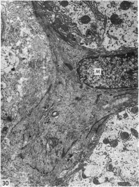
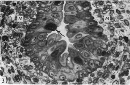
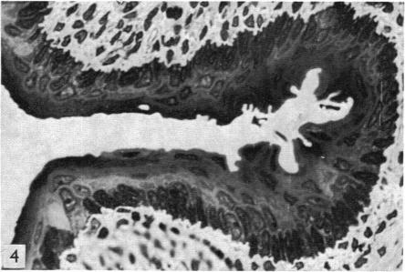
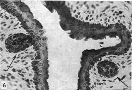
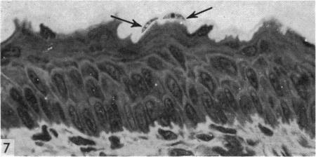
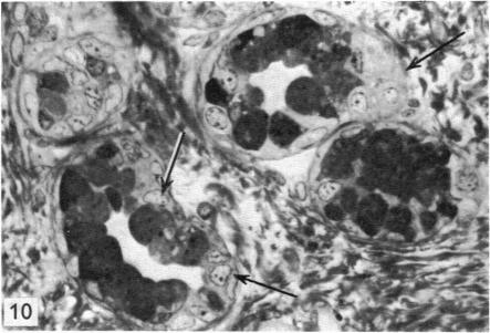
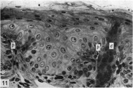
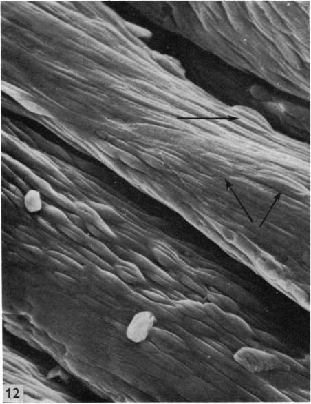
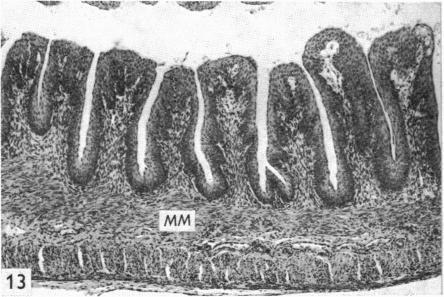
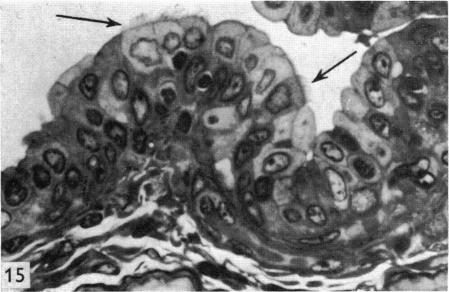
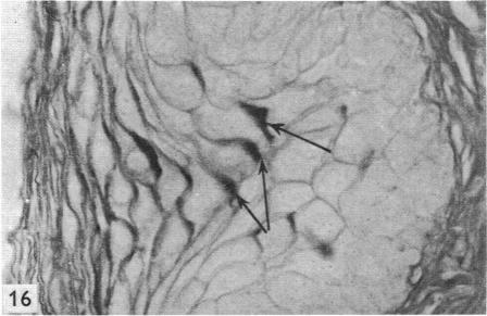
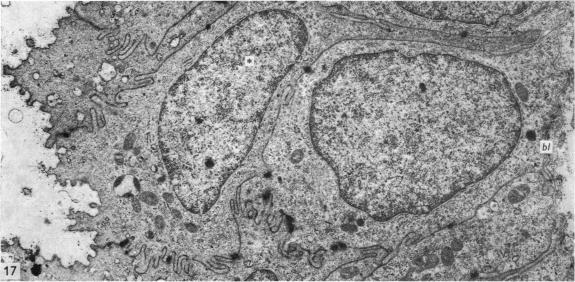
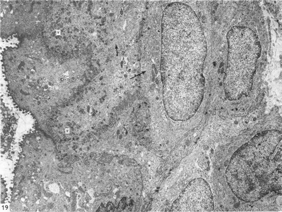
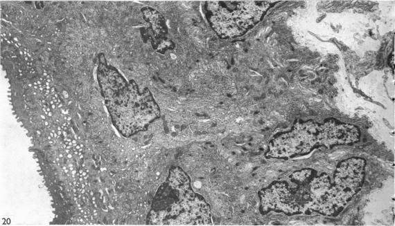
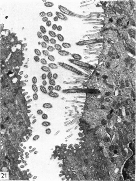
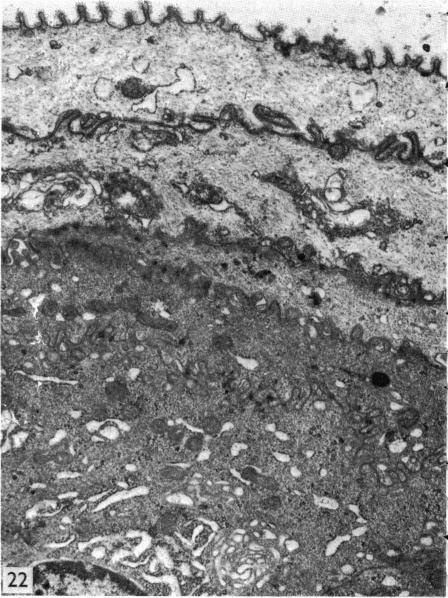
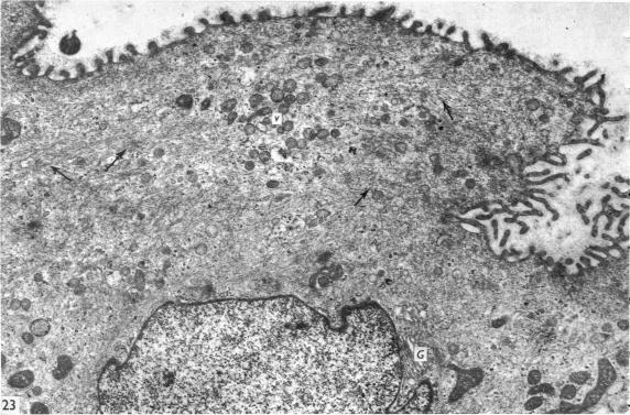
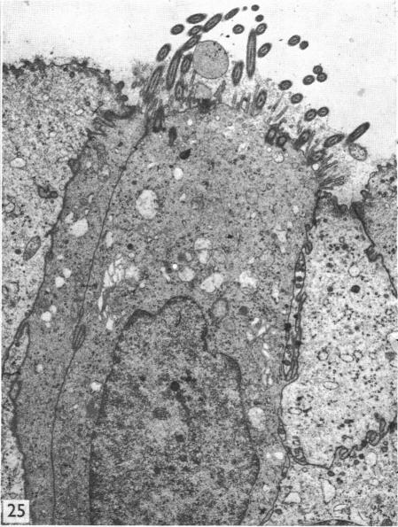
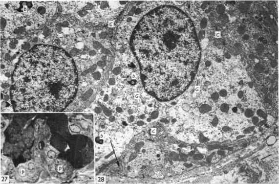
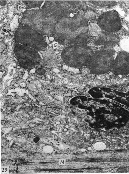
The oesophageal epithelium of the newborn opossum generally is two to three cells in depth and in some regions appears pseudostratified. By the 9th postnatal day the epithelium shows two distinct strata. Ciliated cells and occasional goblet cells also are observed within the epithelium during this stage and in subsequent stages. Cilia persist in the oesophagus of the adult opossum, but are restricted to the depths of the transverse folds found in the distal part of the organ. The epithelium covering the transverse folds of the adult likewise has an immature appearance. By 4-5 cm (ca. 20 days), the epithelium has assumed a more mature appearance and is of greater depth. This and later stages show three basic strata: a germinal layer, a spinous layer and, adjacent to the lumen, a flattened layer of cells that retain their nuclei. The epithelium throughout the postnatal period and in the adult does not undergo complete keratinization. The oesophageal glands begin as outgrowths from the epithelium just prior to 4-5 cm (ca. 20 days). The glands continue their development throughout the remainder of the postnatal period. The secretory units of the oesophageal glands of the the major portion of the secretory elements, and a light, rounded cell type which is less numerous and which occupies the terminal portions of the secretory units. Secretory material of the former appears complex, consisting of both neutral and acid glycoproteins. The secretory product of the light cell type is unknown at present. Both cell types are encompassed by myoepithelial cells. The relationship of the mitotic sequences to the observations made by microscopic examination of the developing oesophagus is discussed.

摘要

新生负鼠的食管上皮通常有两到三层细胞深,在某些区域呈假复层。出生后第9天,上皮显示出两个不同的层。在此阶段及后续阶段,上皮内也观察到纤毛细胞和偶尔的杯状细胞。纤毛在成年负鼠的食管中持续存在,但仅限于器官远端发现的横向褶皱深处。覆盖成年负鼠横向褶皱的上皮同样具有不成熟的外观。到4-5厘米(约20天)时,上皮呈现出更成熟的外观且更深。这个阶段及以后的阶段显示出三个基本层:生发层、棘层,以及与管腔相邻的一层扁平细胞,这些细胞保留着细胞核。在整个出生后时期及成年期,上皮都不会完全角化。食管腺在4-5厘米(约20天)之前开始从上皮长出。这些腺体在出生后剩余时间里继续发育。食管腺的分泌单位主要由大部分分泌成分组成,还有一种浅色、圆形的细胞类型,数量较少,占据分泌单位的末端部分。前者的分泌物质看起来很复杂,由中性和酸性糖蛋白组成。目前尚不清楚浅色细胞类型的分泌产物。两种细胞类型都被肌上皮细胞包围。本文讨论了有丝分裂序列与发育中食管显微镜检查结果之间的关系。

https://cdn.ncbi.nlm.nih.gov/pmc/blobs/82fa/1231902/78bb1e0d4db0/janat00369-0097-c.jpg
https://cdn.ncbi.nlm.nih.gov/pmc/blobs/82fa/1231902/7b1469d734d3/janat00369-0083-a.jpg
https://cdn.ncbi.nlm.nih.gov/pmc/blobs/82fa/1231902/bc145e2b3f42/janat00369-0083-b.jpg
https://cdn.ncbi.nlm.nih.gov/pmc/blobs/82fa/1231902/9fa805309518/janat00369-0083-c.jpg
https://cdn.ncbi.nlm.nih.gov/pmc/blobs/82fa/1231902/8bb9a06ccabe/janat00369-0083-d.jpg
https://cdn.ncbi.nlm.nih.gov/pmc/blobs/82fa/1231902/3a50c1479561/janat00369-0083-e.jpg
https://cdn.ncbi.nlm.nih.gov/pmc/blobs/82fa/1231902/18cde2c51aa6/janat00369-0083-f.jpg
https://cdn.ncbi.nlm.nih.gov/pmc/blobs/82fa/1231902/80418a081bcd/janat00369-0083-g.jpg
https://cdn.ncbi.nlm.nih.gov/pmc/blobs/82fa/1231902/cbed9bbd9521/janat00369-0083-h.jpg
https://cdn.ncbi.nlm.nih.gov/pmc/blobs/82fa/1231902/54d13bc582d2/janat00369-0085-a.jpg
https://cdn.ncbi.nlm.nih.gov/pmc/blobs/82fa/1231902/dda954808a6e/janat00369-0085-b.jpg
https://cdn.ncbi.nlm.nih.gov/pmc/blobs/82fa/1231902/7ee8849f966b/janat00369-0085-c.jpg
https://cdn.ncbi.nlm.nih.gov/pmc/blobs/82fa/1231902/72488076b19f/janat00369-0085-d.jpg
https://cdn.ncbi.nlm.nih.gov/pmc/blobs/82fa/1231902/6a11a959c235/janat00369-0085-e.jpg
https://cdn.ncbi.nlm.nih.gov/pmc/blobs/82fa/1231902/7167db1d39d2/janat00369-0085-f.jpg
https://cdn.ncbi.nlm.nih.gov/pmc/blobs/82fa/1231902/2ec3e44ba652/janat00369-0087-a.jpg
https://cdn.ncbi.nlm.nih.gov/pmc/blobs/82fa/1231902/fc66e52dbdea/janat00369-0087-b.jpg
https://cdn.ncbi.nlm.nih.gov/pmc/blobs/82fa/1231902/45bddc58b592/janat00369-0087-c.jpg
https://cdn.ncbi.nlm.nih.gov/pmc/blobs/82fa/1231902/ce54cc1590ee/janat00369-0087-d.jpg
https://cdn.ncbi.nlm.nih.gov/pmc/blobs/82fa/1231902/855d1b15bb38/janat00369-0091-a.jpg
https://cdn.ncbi.nlm.nih.gov/pmc/blobs/82fa/1231902/02721e56fd68/janat00369-0091-b.jpg
https://cdn.ncbi.nlm.nih.gov/pmc/blobs/82fa/1231902/25a593441759/janat00369-0093-a.jpg
https://cdn.ncbi.nlm.nih.gov/pmc/blobs/82fa/1231902/562ba580272b/janat00369-0093-b.jpg
https://cdn.ncbi.nlm.nih.gov/pmc/blobs/82fa/1231902/ef1469adabe0/janat00369-0093-c.jpg
https://cdn.ncbi.nlm.nih.gov/pmc/blobs/82fa/1231902/15421f513dc2/janat00369-0095-a.jpg
https://cdn.ncbi.nlm.nih.gov/pmc/blobs/82fa/1231902/2eb0a62f6b2e/janat00369-0095-b.jpg
https://cdn.ncbi.nlm.nih.gov/pmc/blobs/82fa/1231902/3f4672012d10/janat00369-0095-c.jpg
https://cdn.ncbi.nlm.nih.gov/pmc/blobs/82fa/1231902/237e63b3668e/janat00369-0097-a.jpg
https://cdn.ncbi.nlm.nih.gov/pmc/blobs/82fa/1231902/5b4f2165b718/janat00369-0097-b.jpg
https://cdn.ncbi.nlm.nih.gov/pmc/blobs/82fa/1231902/78bb1e0d4db0/janat00369-0097-c.jpg

相似文献

1
The postnatal development of the alimentary canal in the opossum. I. Oesophagus.负鼠消化道的产后发育。I. 食管。
J Anat. 1976 Nov;122(Pt 2):293-314.
2
The postnatal development of the alimentary canal in the opossum. III. Small intestine and colon.负鼠消化道的产后发育。III. 小肠和结肠。
J Anat. 1977 Feb;123(Pt 1):21-45.
3
The postnatal development of the alimentary canal in the opossum. II. Stomach.负鼠消化道的出生后发育。II. 胃
J Anat. 1976 Dec;122(Pt 3):499-519.
4
Histology of the esophagus of the adult frog Rana perezi (Anura: Ranidae).成年泽蛙(无尾目:蛙科)食管的组织学
J Morphol. 1992 Jun;212(3):191-200. doi: 10.1002/jmor.1052120302.
5
Studies on the progestational endometrium of the rabbit. II. Electron microscopy, day 0 to day 13 of gonadotrophin-induced pseudopregnancy.兔孕前期子宫内膜的研究。II. 促性腺激素诱导假孕第0天至第13天的电子显微镜观察
Am J Anat. 1975 Mar;142(3):335-65. doi: 10.1002/aja.1001420305.
6
Morphology of differentiating epithelial cells in the respiratory tract of the postnatal opossum (Didelphis virginiana).
Scan Electron Microsc. 1980(3):291-300.
7
Development of esophageal epithelium in the fetal and neonatal mouse.
Anat Rec. 1991 Jun;230(2):225-34. doi: 10.1002/ar.1092300210.
8
Postnatal development and aging of esophageal epithelium in mouse: a light and electron microscopic radioautographic study.
Cell Mol Biol (Noisy-le-grand). 1993 May;39(3):309-16.
9
Development of hamster tracheal epithelium: IV. Cell proliferation and cytodifferentiation in the neonate.
Anat Rec. 1986 Feb;214(2):183-92. doi: 10.1002/ar.1092140213.
10
Structure of the oesophagus and morphometric, histochemical-immunohistochemical profiles of the oesophageal gland during the post-hatching period of Japanese quails (Coturnix coturnix japonica).日本鹌鹑(Coturnix coturnix japonica)孵化后时期食管的结构及食管腺的形态计量学、组织化学 - 免疫组织化学特征
Anat Histol Embryol. 2009 Oct;38(5):330-40. doi: 10.1111/j.1439-0264.2009.00947.x. Epub 2009 Jul 27.

引用本文的文献

1
Genetic and cellular mechanisms regulating anterior foregut and esophageal development.调控前前肠和食管发育的遗传和细胞机制。
Dev Biol. 2012 Sep 1;369(1):54-64. doi: 10.1016/j.ydbio.2012.06.016. Epub 2012 Jun 27.
2
Development of the eye in the North American opossum (Didelphis virginiana).北美负鼠(弗吉尼亚负鼠)眼睛的发育。
J Anat. 1993 Oct;183 ( Pt 2)(Pt 2):343-58.
3
Histochemistry of the striated musculature in the opossum and human oesophagus.负鼠和人类食管横纹肌的组织化学

本文引用的文献

1
Staining of stratified squamous epithelium of mucous membranes and skin of man and monkey by the periodic acid-Schiff method.用高碘酸-希夫氏法对人和猴的粘膜及皮肤的复层鳞状上皮进行染色。
Anat Rec. 1951 Jul;110(3):359-75. doi: 10.1002/ar.1091100307.
2
The normal histology of the esophageal and gastric mucosae of the frog. Rana pipiens.牛蛙食管和胃黏膜的正常组织学
J Exp Zool. 1959 Jun;141:155-73. doi: 10.1002/jez.1401410108.
3
STUDIES ON CILIARY MOVEMENTS: I. MECHANISM OF CILIARY MOVEMENT IN FROG'S OESOPHAGUS.纤毛运动的研究:I. 青蛙食管纤毛运动的机制
J Anat. 1982 Mar;134(Pt 2):407-14.
4
Postnatal development of the epidermis in a marsupial, Didelphis virginiana.有袋动物弗吉尼亚负鼠表皮的产后发育
J Anat. 1978 Jan;125(Pt 1):85-99.
5
Morphological observations on the metanephros in the postnatal opossum, Didelphis virginiana.对产后弗吉尼亚负鼠后肾的形态学观察。
J Anat. 1979 Oct;129(Pt 3):459-77.
6
Morphological observations on the mesonephros in the postnatal opossum, Didelphis virginiana.对产后弗吉尼亚负鼠(Didelphis virginiana)中肾的形态学观察。
J Anat. 1979 Sep;129(Pt 2):377-97.
7
The postnatal development of the alimentary canal in the opossum. III. Small intestine and colon.负鼠消化道的产后发育。III. 小肠和结肠。
J Anat. 1977 Feb;123(Pt 1):21-45.
Arch Int Pharmacodyn Ther. 1964 Aug 1;150:348-55.
4
Developmental changes in the oesophageal epithelium in man.人类食管上皮的发育变化
J Anat. 1952 Oct;86(4):431-42.
5
The histogenesis of the esophagus and crop of the chicken, turkey, guinea fowl and pigeon, with special reference to ciliated epithelium.
Anat Rec. 1952 Oct;114(2):189-211. doi: 10.1002/ar.1091140207.
6
Ultrastructural differentiation of the epithelium and mucous glands of the esophagus in the chick embryo.
J Morphol. 1967 Oct;123(2):121-31. doi: 10.1002/jmor.1051230203.
7
An electron microscopic study of esophageal epithelium in the newborn and adult mouse.新生和成年小鼠食管上皮的电子显微镜研究
Am J Anat. 1967 Sep;121(2):175-95. doi: 10.1002/aja.1001210202.
8
"Membrane-coating" granules of mouse oesophageal and gastric epithelium.
J Invest Dermatol. 1966 Oct;47(4):359-62.
9
On the cytology and cytochemistry of the opossum's bronchial glands.
Am J Anat. 1965 Nov;117(3):311-37. doi: 10.1002/aja.1001170302.
10
Mucin biosynthesis by chick and human oesophagus during ontogenetic metaplasia.鸡和人食管在个体发育化生过程中的黏蛋白生物合成
J Anat. 1970 Jul;107(Pt 1):49-66.