• 文献检索
  • 文档翻译
  • 深度研究
  • 学术资讯
  • Suppr Zotero 插件Zotero 插件
  • 邀请有礼
  • 套餐&价格
  • 历史记录
应用&插件
Suppr Zotero 插件Zotero 插件浏览器插件Mac 客户端Windows 客户端微信小程序
定价
高级版会员购买积分包购买API积分包
服务
文献检索文档翻译深度研究API 文档MCP 服务
关于我们
关于 Suppr公司介绍联系我们用户协议隐私条款
关注我们

Suppr 超能文献

核心技术专利:CN118964589B侵权必究
粤ICP备2023148730 号-1Suppr @ 2026

文献检索

告别复杂PubMed语法,用中文像聊天一样搜索,搜遍4000万医学文献。AI智能推荐,让科研检索更轻松。

立即免费搜索

文件翻译

保留排版,准确专业,支持PDF/Word/PPT等文件格式,支持 12+语言互译。

免费翻译文档

深度研究

AI帮你快速写综述,25分钟生成高质量综述,智能提取关键信息,辅助科研写作。

立即免费体验

负鼠消化道的产后发育。III. 小肠和结肠。

The postnatal development of the alimentary canal in the opossum. III. Small intestine and colon.

作者信息

Krause W J, Cutts J H, Leeson C R

出版信息

J Anat. 1977 Feb;123(Pt 1):21-45.

PMID:190199
原文链接:https://pmc.ncbi.nlm.nih.gov/articles/PMC1234251/
Abstract

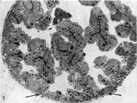
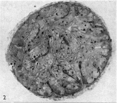
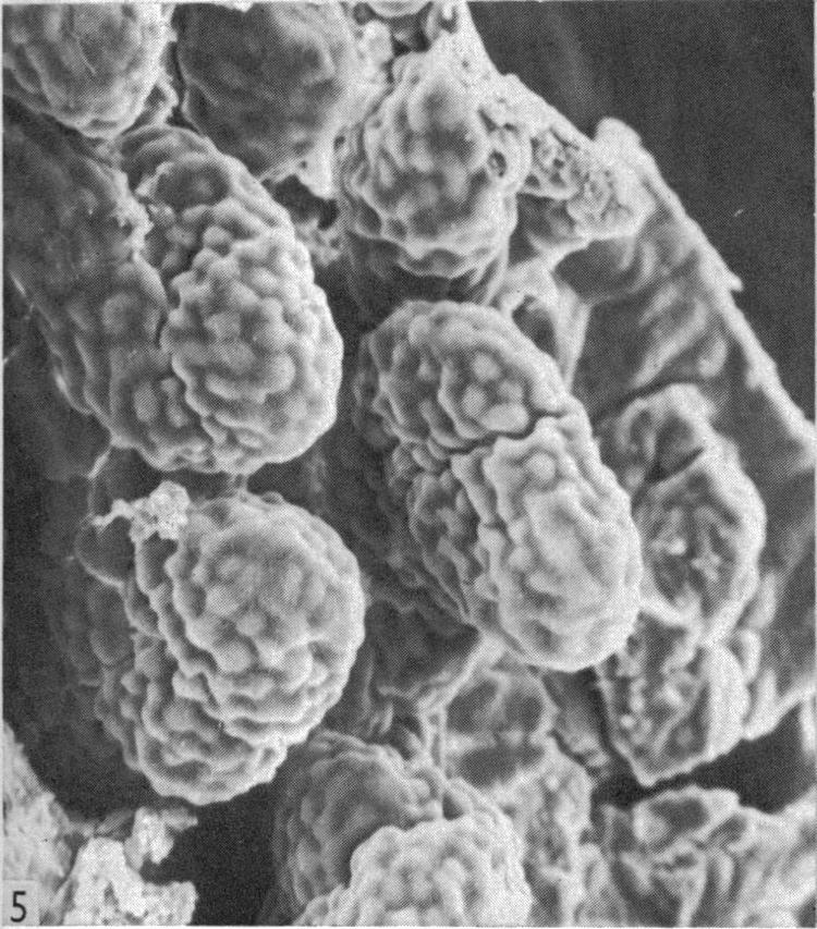
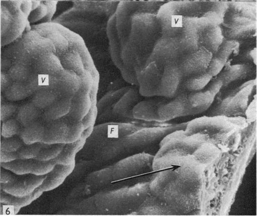
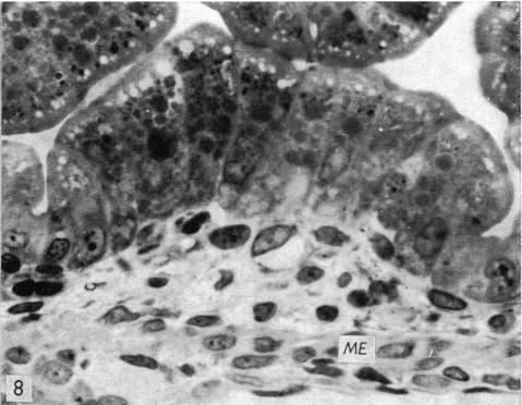
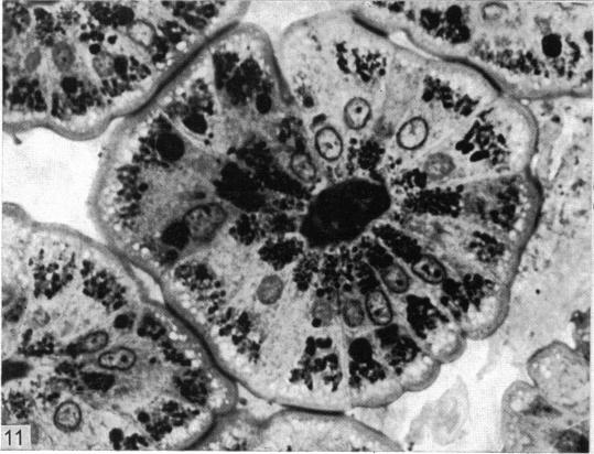
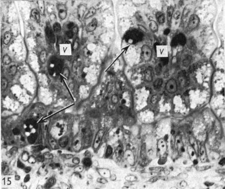
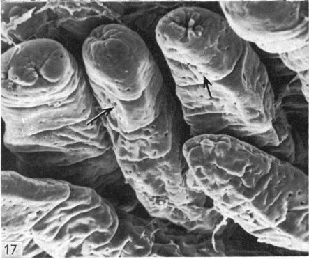
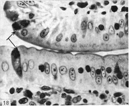
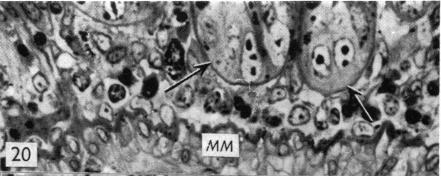
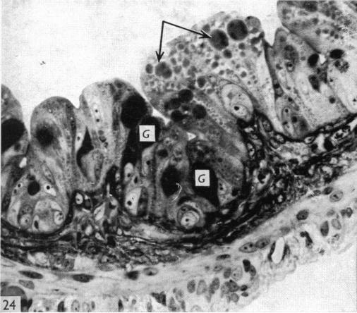
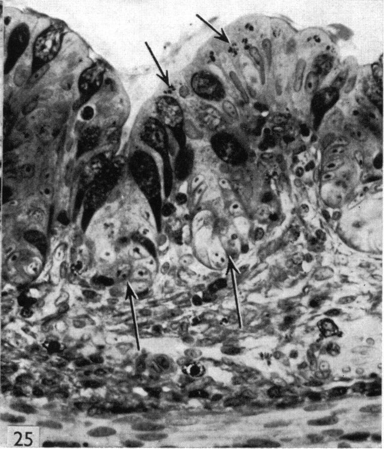
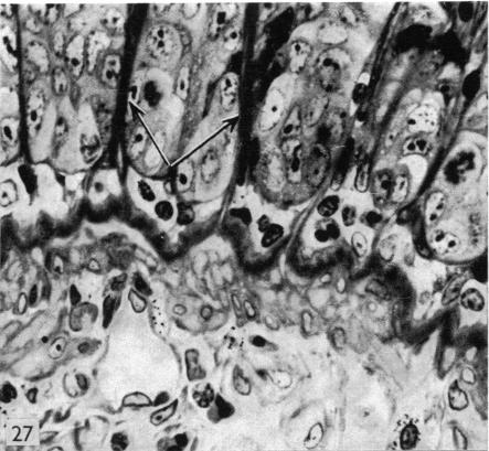
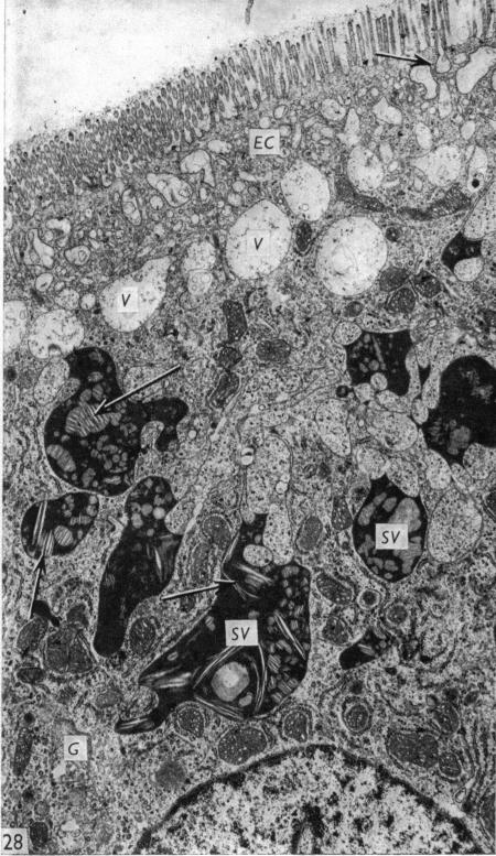
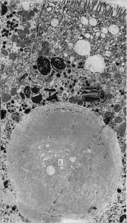
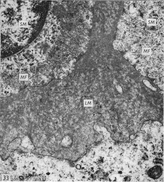
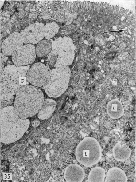
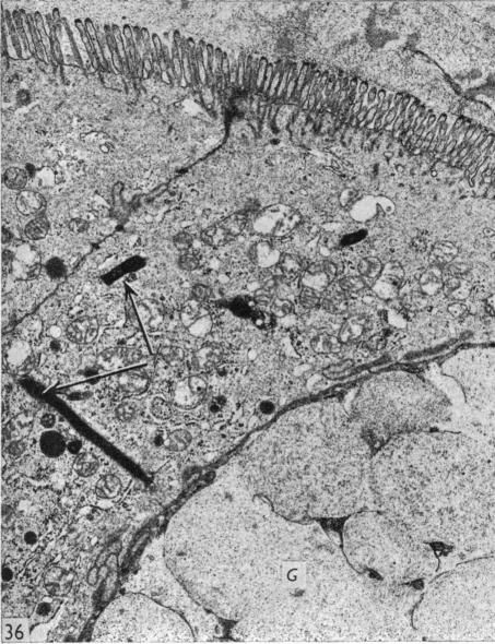
The duodenum of the newborn opossum exhibits a patent lumen containing scattered elongate villi, whereas the distal segments of the small intestine are smaller in diameter and are filled with short immature villi. The muscularis externa through the small intestine consists of a single layer of myoblasts. Interposed between the intestinal lining epithelium and the muscularis externa is an extensive capillary bed that occupies a considerable proportion of the intestinal wall. Additional villi appear to form during the postnatal period as a result of evaginations of the epithelium, together with underlying connective tissue and vasculature, into the intestinal lumen. Intestinal glands are not observed until 8.5cm, and are shallow in depth even in the adult. The epithelium of the entire small intestine is modified for absorption until just prior to weaning. The principal intestinal lining cells show an extensive apical endocytic complex, large supranuclear vacuoles and numerous cytoplasmic inclusions. Intestinal epithelial cells of the colon also appear to be modified for absorption during the first two weeks after birth. Although goblet cells and Paneth cells are present during the suckling period, they do not comprise a significant population in the intestinal epithelium until after weaning. In contrast to the small intestine, goblet cells are numerous in the colon by the ninth postnatal day. The significance of macromolecular absorption and the possibility of passive immunity being transmitted in the opossum during suckling are discussed and related to similar events that occur in the slckling young of several eutherian species. The possible functional significance of two large membranes that develop in the lamina propria of the intestines after weaning also is discussed.

摘要

新生负鼠的十二指肠呈现出一个管腔通畅的结构,其中含有散在的细长绒毛,而小肠的远端部分直径较小,充满了短小的未成熟绒毛。贯穿小肠的外肌层由单层成肌细胞组成。在肠内衬上皮和外肌层之间是一个广泛的毛细血管床,它占据了肠壁相当大的比例。出生后,由于上皮连同其下方的结缔组织和脉管系统向肠腔内突出,额外的绒毛似乎开始形成。直到肠长达到8.5厘米时才观察到肠腺,即使在成年个体中肠腺也很浅。在断奶前,整个小肠的上皮都经过修饰以进行吸收。主要的肠内衬细胞显示出广泛的顶端内吞复合体、大的核上空泡和众多的细胞质内含物。结肠的肠上皮细胞在出生后的头两周似乎也经过修饰以进行吸收。尽管在哺乳期间存在杯状细胞和潘氏细胞,但直到断奶后它们在肠上皮中才占显著比例。与小肠不同,到出生后第九天结肠中的杯状细胞就很多了。本文讨论了负鼠在哺乳期间大分子吸收的意义以及被动免疫传递的可能性,并将其与几种真兽类物种的哺乳幼崽中发生的类似事件相关联。还讨论了断奶后在肠固有层中形成的两个大膜可能具有的功能意义。

https://cdn.ncbi.nlm.nih.gov/pmc/blobs/4bdd/1234251/e30baba97a47/janat00365-0044-d.jpg
https://cdn.ncbi.nlm.nih.gov/pmc/blobs/4bdd/1234251/3e90e02b3d22/janat00365-0028-a.jpg
https://cdn.ncbi.nlm.nih.gov/pmc/blobs/4bdd/1234251/528a2691c0c4/janat00365-0028-b.jpg
https://cdn.ncbi.nlm.nih.gov/pmc/blobs/4bdd/1234251/80fc96c810b6/janat00365-0028-c.jpg
https://cdn.ncbi.nlm.nih.gov/pmc/blobs/4bdd/1234251/8e62da9790a3/janat00365-0028-d.jpg
https://cdn.ncbi.nlm.nih.gov/pmc/blobs/4bdd/1234251/99776a1da285/janat00365-0028-e.jpg
https://cdn.ncbi.nlm.nih.gov/pmc/blobs/4bdd/1234251/c50f60de58ef/janat00365-0028-f.jpg
https://cdn.ncbi.nlm.nih.gov/pmc/blobs/4bdd/1234251/21b9bfe581a3/janat00365-0030-a.jpg
https://cdn.ncbi.nlm.nih.gov/pmc/blobs/4bdd/1234251/d455e16c9586/janat00365-0030-b.jpg
https://cdn.ncbi.nlm.nih.gov/pmc/blobs/4bdd/1234251/2c7247880b8e/janat00365-0030-c.jpg
https://cdn.ncbi.nlm.nih.gov/pmc/blobs/4bdd/1234251/2c4df70e15b6/janat00365-0030-d.jpg
https://cdn.ncbi.nlm.nih.gov/pmc/blobs/4bdd/1234251/3ea17e12fd03/janat00365-0030-e.jpg
https://cdn.ncbi.nlm.nih.gov/pmc/blobs/4bdd/1234251/628b7614e827/janat00365-0030-f.jpg
https://cdn.ncbi.nlm.nih.gov/pmc/blobs/4bdd/1234251/8e3aa1166261/janat00365-0032-a.jpg
https://cdn.ncbi.nlm.nih.gov/pmc/blobs/4bdd/1234251/97d1ce48a344/janat00365-0032-b.jpg
https://cdn.ncbi.nlm.nih.gov/pmc/blobs/4bdd/1234251/3f42ddaff7d7/janat00365-0032-c.jpg
https://cdn.ncbi.nlm.nih.gov/pmc/blobs/4bdd/1234251/7d9443e6e707/janat00365-0032-d.jpg
https://cdn.ncbi.nlm.nih.gov/pmc/blobs/4bdd/1234251/d45a3e0828b9/janat00365-0032-e.jpg
https://cdn.ncbi.nlm.nih.gov/pmc/blobs/4bdd/1234251/27757f4be4f5/janat00365-0032-f.jpg
https://cdn.ncbi.nlm.nih.gov/pmc/blobs/4bdd/1234251/0d5dc571ea4f/janat00365-0034-a.jpg
https://cdn.ncbi.nlm.nih.gov/pmc/blobs/4bdd/1234251/53542ad734f6/janat00365-0034-b.jpg
https://cdn.ncbi.nlm.nih.gov/pmc/blobs/4bdd/1234251/07e3dd6df827/janat00365-0034-c.jpg
https://cdn.ncbi.nlm.nih.gov/pmc/blobs/4bdd/1234251/fda0f5307d76/janat00365-0034-d.jpg
https://cdn.ncbi.nlm.nih.gov/pmc/blobs/4bdd/1234251/1fcdd93494fe/janat00365-0034-e.jpg
https://cdn.ncbi.nlm.nih.gov/pmc/blobs/4bdd/1234251/c0e0cea3d55a/janat00365-0034-f.jpg
https://cdn.ncbi.nlm.nih.gov/pmc/blobs/4bdd/1234251/1d24e21f4ea4/janat00365-0034-g.jpg
https://cdn.ncbi.nlm.nih.gov/pmc/blobs/4bdd/1234251/2f6febb0545b/janat00365-0038-a.jpg
https://cdn.ncbi.nlm.nih.gov/pmc/blobs/4bdd/1234251/e13b795fda86/janat00365-0038-b.jpg
https://cdn.ncbi.nlm.nih.gov/pmc/blobs/4bdd/1234251/b78c69b057e2/janat00365-0038-c.jpg
https://cdn.ncbi.nlm.nih.gov/pmc/blobs/4bdd/1234251/c75bc1abea36/janat00365-0038-d.jpg
https://cdn.ncbi.nlm.nih.gov/pmc/blobs/4bdd/1234251/3dbcf34fff2e/janat00365-0042-a.jpg
https://cdn.ncbi.nlm.nih.gov/pmc/blobs/4bdd/1234251/cdc146112a6c/janat00365-0042-b.jpg
https://cdn.ncbi.nlm.nih.gov/pmc/blobs/4bdd/1234251/38337b3cc704/janat00365-0042-c.jpg
https://cdn.ncbi.nlm.nih.gov/pmc/blobs/4bdd/1234251/88a367e3df39/janat00365-0042-d.jpg
https://cdn.ncbi.nlm.nih.gov/pmc/blobs/4bdd/1234251/beb48805a381/janat00365-0044-a.jpg
https://cdn.ncbi.nlm.nih.gov/pmc/blobs/4bdd/1234251/7c6afc0cf083/janat00365-0044-b.jpg
https://cdn.ncbi.nlm.nih.gov/pmc/blobs/4bdd/1234251/2a73778af109/janat00365-0044-c.jpg
https://cdn.ncbi.nlm.nih.gov/pmc/blobs/4bdd/1234251/e30baba97a47/janat00365-0044-d.jpg

相似文献

1
The postnatal development of the alimentary canal in the opossum. III. Small intestine and colon.负鼠消化道的产后发育。III. 小肠和结肠。
J Anat. 1977 Feb;123(Pt 1):21-45.
2
Enteroendocrine cells in the developing opossum small intestine and colon.发育中的负鼠小肠和结肠中的肠内分泌细胞。
J Anat. 1989 Feb;162:83-96.
3
The postnatal development of the alimentary canal in the opossum. II. Stomach.负鼠消化道的出生后发育。II. 胃
J Anat. 1976 Dec;122(Pt 3):499-519.
4
Prenatal and postnatal development of the small intestine in the insectivore Suncus murinus.食虫动物臭鼩小肠的产前和产后发育
Am J Anat. 1988 Sep;183(1):57-67. doi: 10.1002/aja.1001830104.
5
Structural and mechanical architecture of the intestinal villi and crypts in the rat intestine: integrative reevaluation from ultrastructural analysis.大鼠肠道绒毛和隐窝的结构与力学架构:基于超微结构分析的综合重新评估
Anat Embryol (Berl). 2005 Aug;210(1):1-12. doi: 10.1007/s00429-005-0011-y. Epub 2005 Jul 26.
6
The postnatal development of the alimentary canal in the opossum. I. Oesophagus.负鼠消化道的产后发育。I. 食管。
J Anat. 1976 Nov;122(Pt 2):293-314.
7
Some morphological and histochemical studies on the intestinal tract of the Brazilian sloth (Bradypus tridactylus).关于巴西树懒(三趾树懒)肠道的一些形态学和组织化学研究。
Gegenbaurs Morphol Jahrb. 1989;135(2):367-77.
8
Selective loss of sialic acid from rat small intestinal epithelial cells during postnatal development: demonstration with lectin-gold techniques.
Eur J Cell Biol. 1990 Dec;53(2):255-66.
9
Light and scanning electron microscopy evaluation of the postnatal small intestinal mucosa development in pigs.仔猪出生后小肠黏膜发育的光镜和扫描电镜评估
J Physiol Pharmacol. 2005 Jun;56 Suppl 3:71-87.
10
Scanning electron, light, and transmission electron microscopy of intestine of gnotobiotic calf.无菌小牛肠道的扫描电子显微镜、光学显微镜和透射电子显微镜检查
Am J Vet Res. 1975 Jul;36(7):985-93.

引用本文的文献

1
A quantitative study of the morphological development and bacterial colonisation of the gut of the tammar wallaby Macropus eugenii eugenii and brushtail possum Trichosurus vulpecula during in-pouch development.对袋貂(Macropus eugenii eugenii)和帚尾袋貂(Trichosurus vulpecula)育儿袋内发育期间肠道形态发育和细菌定植的定量研究。
J Comp Physiol B. 2006 Nov;176(8):763-74. doi: 10.1007/s00360-006-0097-4. Epub 2006 Jul 4.
2
Development of the eye in the North American opossum (Didelphis virginiana).北美负鼠(弗吉尼亚负鼠)眼睛的发育。
J Anat. 1993 Oct;183 ( Pt 2)(Pt 2):343-58.
3
Leukocytes in the peripheral blood of the developing opossum.

本文引用的文献

1
SOME ELECTRON MICROSCOPIC OBSERVATIONS ON THE LAMINA PROPRIA OF THE GUT, WITH COMMENTS ON THE CLOSE ASSOCIATION OF MACROPHAGES, PLASMA CELLS, AND EOSINOPHILS.肠道固有层的一些电子显微镜观察,兼论巨噬细胞、浆细胞和嗜酸性粒细胞的紧密关联
Anat Rec. 1964 Jul;149:453-73. doi: 10.1002/ar.1091490315.
2
AN ELECTRON MICROSCOPE STUDY OF MATURE AND DIFFERENTIATING PANETH CELLS IN THE RAT, ESPECIALLY OF THEIR ENDOPLASMIC RETICULUM AND LYSOSOMES.大鼠成熟和分化中的潘氏细胞的电子显微镜研究,特别是对其内质网和溶酶体的研究。
J Cell Biol. 1964 Sep;22(3):633-52. doi: 10.1083/jcb.22.3.633.
3
SPECIFIC STAINING OF SULPHATE GROUPS WITH ALCIAN BLUE AT LOW PH.
发育中的负鼠外周血中的白细胞。
J Anat. 1980 Jan;130(Pt 1):113-20.
4
The development of the stomach in clarias lazera and the intestinal absorption of protein macromolecules.胡子鲶胃的发育及蛋白质大分子的肠道吸收
Cell Tissue Res. 1981;215(2):397-415. doi: 10.1007/BF00239123.
5
A scanning and transmission electron microscopical study of the morphogenesis of human colonic villi.人结肠绒毛形态发生的扫描电镜与透射电镜研究
Anat Embryol (Berl). 1982 Dec;165(3):437-55. doi: 10.1007/BF00305579.
6
Quantitative distribution of enteroendocrine cells in the gastrointestinal tract of the adult opossum, Didelphis virginiana.成年弗吉尼亚负鼠(Didelphis virginiana)胃肠道中肠内分泌细胞的定量分布。
J Anat. 1985 Jun;140 ( Pt 4)(Pt 4):591-605.
7
Enteroendocrine cells in the developing opossum small intestine and colon.发育中的负鼠小肠和结肠中的肠内分泌细胞。
J Anat. 1989 Feb;162:83-96.
8
Postnatal development and differentiation of the opossum submandibular gland.负鼠下颌下腺的出生后发育与分化
J Anat. 1978 Jun;126(Pt 2):329-51.
9
Scanning electron microscopic studies of ileal epithelial cells in suckling rats.哺乳大鼠回肠上皮细胞的扫描电子显微镜研究。
Anat Embryol (Berl). 1978 May 31;153(1):1-8. doi: 10.1007/BF00569845.
10
Postnatal development of the epidermis in a marsupial, Didelphis virginiana.有袋动物弗吉尼亚负鼠表皮的产后发育
J Anat. 1978 Jan;125(Pt 1):85-99.
低pH值条件下阿尔辛蓝对硫酸基团的特异性染色
J Histochem Cytochem. 1964 Apr;12:309. doi: 10.1177/12.4.309.
4
PROTEIN SYNTHESIS, STORAGE, AND DISCHARGE IN THE PANCREATIC EXOCRINE CELL. AN AUTORADIOGRAPHIC STUDY.胰腺外分泌细胞中的蛋白质合成、储存与释放。一项放射自显影研究。
J Cell Biol. 1964 Mar;20(3):473-95. doi: 10.1083/jcb.20.3.473.
5
STUDIES ON SMALL INTESTINAL CRYPT EPITHELIUM. I. THE FINE STRUCTURE OF THE CRYPT EPITHELIUM OF THE PROXIMAL SMALL INTESTINE OF FASTING HUMANS.小肠隐窝上皮的研究。I. 禁食人类近端小肠隐窝上皮的精细结构
J Cell Biol. 1963 Sep;18(3):599-620. doi: 10.1083/jcb.18.3.599.
6
Synthesis and migration of proteins in the cells of the exocrine pancreas as revealed by specific activity determination from radioautographs.通过放射自显影片的比活性测定揭示外分泌胰腺细胞中蛋白质的合成与迁移
J Cell Biol. 1963 Jan;16(1):1-24. doi: 10.1083/jcb.16.1.1.
7
A cytochemical analysis of Paneth cell secretion in the mouse.
Anat Rec. 1961 May;140:17-22. doi: 10.1002/ar.1091400103.
8
Preliminary observations of the Paneth cells of the Tamandua tetradactyla Lin.四指食蚁兽潘氏细胞的初步观察
Acta Anat (Basel). 1959;38:345-52. doi: 10.1159/000141538.
9
The ingestion of proteins and colloidal materials by columnar absorptive cells of the small intestine in suckling rats and mice.乳鼠和幼鼠小肠柱状吸收细胞对蛋白质和胶体物质的摄取。
J Biophys Biochem Cytol. 1959 Jan 25;5(1):41-50. doi: 10.1083/jcb.5.1.41.
10
Histological and histochemical observations on the intestinal cells of lambs and kids absorbing colostrum.对摄取初乳的羔羊和幼山羊肠道细胞的组织学和组织化学观察。
Nature. 1956 Dec 15;178(4546):1353-4. doi: 10.1038/1781353b0.