• 文献检索
  • 文档翻译
  • 深度研究
  • 学术资讯
  • Suppr Zotero 插件Zotero 插件
  • 邀请有礼
  • 套餐&价格
  • 历史记录
应用&插件
Suppr Zotero 插件Zotero 插件浏览器插件Mac 客户端Windows 客户端微信小程序
定价
高级版会员购买积分包购买API积分包
服务
文献检索文档翻译深度研究API 文档MCP 服务
关于我们
关于 Suppr公司介绍联系我们用户协议隐私条款
关注我们

Suppr 超能文献

核心技术专利:CN118964589B侵权必究
粤ICP备2023148730 号-1Suppr @ 2026

文献检索

告别复杂PubMed语法,用中文像聊天一样搜索,搜遍4000万医学文献。AI智能推荐,让科研检索更轻松。

立即免费搜索

文件翻译

保留排版,准确专业,支持PDF/Word/PPT等文件格式,支持 12+语言互译。

免费翻译文档

深度研究

AI帮你快速写综述,25分钟生成高质量综述,智能提取关键信息,辅助科研写作。

立即免费体验

小鼠腺垂体中的细胞分化和门静脉血管发育。

Cyto-differentiation and portal vascular development in the mouse adenohypophysis.

作者信息

Dearden N M, Holmes R L

出版信息

J Anat. 1976 Jul;121(Pt 3):551-69.

PMID:1018008
原文链接:https://pmc.ncbi.nlm.nih.gov/articles/PMC1231746/
Abstract

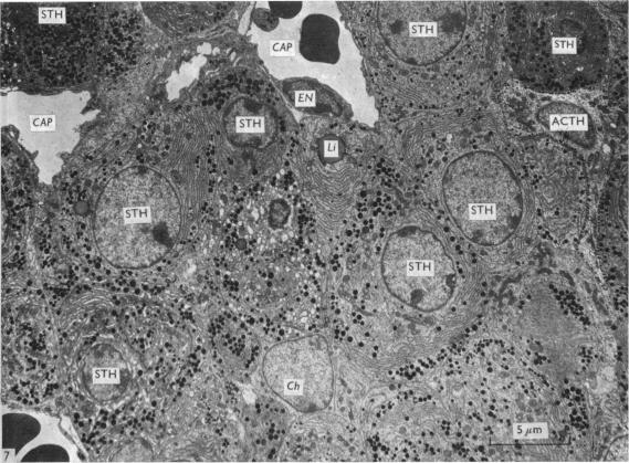
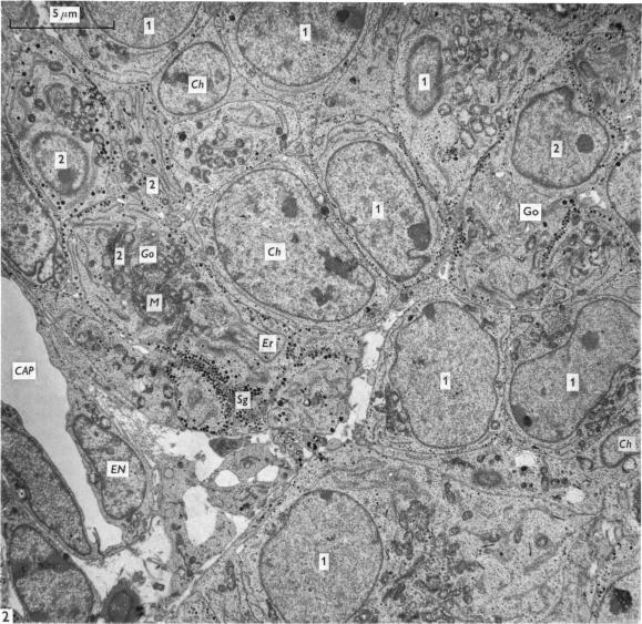
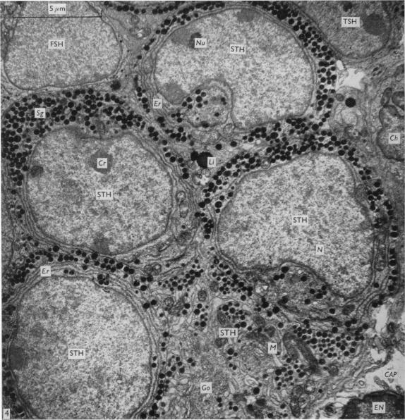
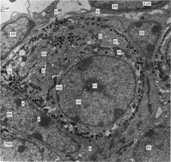
Pituitaries of fetal and postnatal (15 days p.c.-28 days p.n.) and adult (male) mice were studied by light and electron microscopy to correlate the developmental pattern of the hypothalamo-hypophysial vascular system with the time of onset of function of the adenohypophysis. The superior and anterior regions of the adenohypophysis become vascularized at 17 days p.c., when portal vessels extend from oral primary plexus to the pars distalis for the first time. Adenohypophysial vascularity and the number of portal vessels steadily increase to reach the adult pattern at 5 days p.n. At 1 day p.n. deep capillary loops appear in the caudal regions of the oral primary plexus; a capillary (tangential) plexus underlies the ependymal lining of the third ventricle by 6 days p.n. Superficial capillary loops were not observed until the third postnatal week. Granulation of secretory cells commences at 16 days p.c., predominantly in the upper and anterior adenohypophysis; at 17 days thyrotropes, gonadotropes and corticotropes are recognizable and by morphological criteria appear actively secretory on days 17-18 p.c., although few appear active at 19 days p.c. and 1 day p.n. Somatotropes are first seen at 18 days p.c., predominantly in the central and lateral regions of the pars distalis. Active secretory cells increase in number over the period 2-10 days p.n., but after 11 days p.n. thyrotropes and corticotropes seem to become progressively less active; fewer gonadotropes are seen after 15 days p.n., and these apparently become progressively less active from day 19. Most somatotropes appear active until 28 days p.n. The observations suggest that hypothalamic control of adenohypophysial function may exist in the mouse from 17 days p.c.

摘要

运用光镜和电镜技术,对胎鼠、出生后(妊娠15天至出生后28天)及成年(雄性)小鼠的垂体进行了研究,以探寻下丘脑 - 垂体血管系统的发育模式与腺垂体功能起始时间之间的关联。在妊娠17天时,腺垂体的上部和前部开始血管化,此时门静脉首次从口腔初级丛延伸至远侧部。腺垂体的血管化程度和门静脉数量持续稳定增加,至出生后5天时达到成年模式。出生后1天,口腔初级丛的尾部区域出现深部毛细血管袢;到出生后6天,第三脑室室管膜下衬有毛细血管(切线状)丛。直到出生后第三周才观察到浅表毛细血管袢。分泌细胞的颗粒化在妊娠16天时开始,主要发生在腺垂体的上部和前部;17天时,促甲状腺激素细胞、促性腺激素细胞和促肾上腺皮质激素细胞可被识别,从形态学标准来看,在妊娠17 - 18天时这些细胞表现为活跃分泌状态,不过在妊娠19天和出生后1天时仅有少数细胞呈活跃状态。生长激素细胞在妊娠18天时首次出现,主要位于远侧部的中央和外侧区域。在出生后2 - 10天期间,活跃分泌细胞的数量增加,但出生后11天之后,促甲状腺激素细胞和促肾上腺皮质激素细胞的活性似乎逐渐降低;出生后15天之后,促性腺激素细胞数量减少,且从第19天起其活性明显逐渐降低。大多数生长激素细胞在出生后28天前都保持活跃状态。这些观察结果表明,在小鼠中,从妊娠17天起可能就存在下丘脑对腺垂体功能的控制。

https://cdn.ncbi.nlm.nih.gov/pmc/blobs/4ad8/1231746/c88a93ad6207/janat00373-0123-a.jpg
https://cdn.ncbi.nlm.nih.gov/pmc/blobs/4ad8/1231746/a91889e4724e/janat00373-0111-a.jpg
https://cdn.ncbi.nlm.nih.gov/pmc/blobs/4ad8/1231746/928f49a8ec2e/janat00373-0116-a.jpg
https://cdn.ncbi.nlm.nih.gov/pmc/blobs/4ad8/1231746/36d015b1152f/janat00373-0118-a.jpg
https://cdn.ncbi.nlm.nih.gov/pmc/blobs/4ad8/1231746/355a06b4b1f4/janat00373-0120-a.jpg
https://cdn.ncbi.nlm.nih.gov/pmc/blobs/4ad8/1231746/bd21eb9305e1/janat00373-0121-a.jpg
https://cdn.ncbi.nlm.nih.gov/pmc/blobs/4ad8/1231746/ac3424e42d90/janat00373-0122-a.jpg
https://cdn.ncbi.nlm.nih.gov/pmc/blobs/4ad8/1231746/c88a93ad6207/janat00373-0123-a.jpg

相似文献

1
Cyto-differentiation and portal vascular development in the mouse adenohypophysis.小鼠腺垂体中的细胞分化和门静脉血管发育。
J Anat. 1976 Jul;121(Pt 3):551-69.
2
Early development of the pituitary gland in Acipenser naccarii (Chondrostei, Acipenseriformes): an immunocytochemical study.纳氏鲟(软骨硬鳞鱼纲,鲟形目)垂体腺的早期发育:一项免疫细胞化学研究
Anat Embryol (Berl). 2004 Jul;208(4):311-21. doi: 10.1007/s00429-004-0402-5. Epub 2004 Jul 2.
3
Anterior and posterior groups of portal vessels in the avian pituitary: incidence in forty nine species.鸟类垂体门脉血管的前后组:49种鸟类中的发生率
Arch Anat Microsc Morphol Exp. 1975;64(4):359-74.
4
Scanning microscopy of pituitary vascular casts.垂体血管铸型的扫描显微镜检查。
Am J Anat. 1976 Jul;146(3):273-301. doi: 10.1002/aja.1001460305.
5
The hypothalamo-hypophysial vascular relationship in Indian fresh-water goby, Glossogobius giuris (Ham.).920u.印度淡水虾虎鱼(Glossogobius giuris (Ham.))下丘脑 - 垂体的血管关系。920u。
Gegenbaurs Morphol Jahrb. 1975;121(1):55-76.
6
Gene expression of vascular endothelial growth factor-A in the pituitary during formation of the vascular system in the hypothalamic-pituitary axis of the rat.大鼠下丘脑 - 垂体轴血管系统形成过程中垂体中血管内皮生长因子 -A 的基因表达
Cell Tissue Res. 2006 Apr;324(1):87-95. doi: 10.1007/s00441-005-0115-y. Epub 2006 Jan 13.
7
Hypothalamo-hypophysial vascularization of the teleost, Puntius sophore (Ham.) with special reference to its tetrapodan features.印度波鱼(哈姆)硬骨鱼下丘脑 - 垂体的血管形成,特别涉及其四足动物特征。
Anat Anz. 1978;143(1):110-9.
8
Morphological, developmental and immunohistochemical observations on the opossum pituitary with emphasis on the pars intermedia.负鼠垂体的形态学、发育学及免疫组织化学观察,重点关注垂体中间部。
Acta Histochem. 1990;89(1):37-56. doi: 10.1016/S0065-1281(11)80312-0.
9
Ultrastructural study of the development of the pars distalis (anterior pituitary) in the foal.马驹远侧部(垂体前叶)发育的超微结构研究。
J Reprod Fertil Suppl. 1982;32:583-8.
10
Structure of the neurohypophysis and the hypothalamo-hypophysial vascularization in the teleost Channa punctatus Bloch.斑点鳢(Channa punctatus Bloch)神经垂体的结构及下丘脑 - 垂体血管形成
Z Mikrosk Anat Forsch. 1975;89(5):824-38.

引用本文的文献

1
Epithelial cell integrin β1 is required for developmental angiogenesis in the pituitary gland.垂体发育性血管生成需要上皮细胞整合素β1。
Proc Natl Acad Sci U S A. 2016 Nov 22;113(47):13408-13413. doi: 10.1073/pnas.1614970113. Epub 2016 Nov 3.
2
Cold-shock-domain protein A (CSDA) contributes posttranscriptionally to gonadotropin-releasing hormone-regulated expression of Egr1 and indirectly to Lhb.冷休克域蛋白 A(CSDA)在后转录水平上有助于促性腺激素释放激素调节 Egr1 的表达,并间接地有助于 Lhb 的表达。
Biol Reprod. 2012 Feb 29;86(2):53. doi: 10.1095/biolreprod.111.093658. Print 2012 Feb.
3
Postnatal development of somatotrophs and mammotrophs in the pars distalis of the C57BL mouse.

本文引用的文献

1
The site of thyrotrophin and gonadotrophin production in the rat pituitary studied by McManus-Hotchkiss staining for glycoprotein.用麦克马纳斯-霍奇基斯糖蛋白染色法研究大鼠垂体中促甲状腺激素和促性腺激素的产生部位。
Endocrinology. 1951 Aug;49(2):244-64. doi: 10.1210/endo-49-2-244.
2
The use of lead citrate at high pH as an electron-opaque stain in electron microscopy.在电子显微镜检查中,将高pH值的柠檬酸铅用作电子不透明染色剂。
J Cell Biol. 1963 Apr;17(1):208-12. doi: 10.1083/jcb.17.1.208.
3
Isolation of rat pituitary granules and the study of their biochemical properties and hormonal activities.
Cell Tissue Res. 1980;211(3):441-8. doi: 10.1007/BF00234398.
4
Patterns of proliferation in the hypophysis of the mouse embryo. A quantitative autoradiographic study.小鼠胚胎垂体中的增殖模式。一项定量放射自显影研究。
Anat Embryol (Berl). 1980;159(1):101-13. doi: 10.1007/BF00299259.
5
Correlative ontogenetic development of catecholamine- and LHRH-containing nerve endings in the median eminence of the rat.大鼠正中隆起中含儿茶酚胺和促性腺激素释放激素的神经末梢的相关个体发生发育
Cell Tissue Res. 1981;216(1):31-8. doi: 10.1007/BF00234542.
6
Structure of the pars distalis in pouch-young tammar wallabies (Macropus eugenii).育儿袋幼龄帚尾袋貂(尤金袋鼠)远侧部的结构。
Cell Tissue Res. 1983;230(3):587-603. doi: 10.1007/BF00216203.
7
Structure of the pars distalis in the adult tammar wallaby (Macropus eugenii).成年帚尾袋鼩(短尾矮袋鼠)远侧部的结构
Cell Tissue Res. 1983;229(1):155-74. doi: 10.1007/BF00217888.
8
Ontogeny of gonadotropic and thyrotropic cells in fetal mouse anterior pituitary. Comparison between two species C57 BL6 and Balb/C.胎鼠垂体前叶促性腺细胞和促甲状腺细胞的个体发生。两种品系C57 BL6和Balb/C的比较。
Anat Embryol (Berl). 1988;178(1):21-7. doi: 10.1007/BF00305010.
9
The ultrastructure of the pituitary and the adrenal gland of three newborn marsupials (Dasyurus hallucatus, Trichosurus vulpecula and Isoodon macrourus).三种有袋类新生动物(袋鼬、帚尾袋貂和长吻袋狸)垂体和肾上腺的超微结构
Anat Embryol (Berl). 1988;177(5):395-402. doi: 10.1007/BF00304736.
10
Immunofluorescent analysis of somatotroph distribution in the adenohypophysis of developing lit/lit mice.发育中的lit/lit小鼠腺垂体中生长激素细胞分布的免疫荧光分析。
J Anat. 1988 Feb;156:51-9.
大鼠垂体颗粒的分离及其生化特性和激素活性的研究。
J Cell Biol. 1963 Apr;17(1):67-86. doi: 10.1083/jcb.17.1.67.
4
Hypophysiotrophic area in the hypothalamus.下丘脑的促垂体区。
J Endocrinol. 1962 Oct;25:147-54. doi: 10.1677/joe.0.0250147.
5
A postnatal histogenetic study of the anterior pituitary of the mouse.小鼠垂体前叶的产后组织发生学研究。
Okajimas Folia Anat Jpn. 1957 Aug;30(2-3):177-95. doi: 10.2535/ofaj1936.30.2-3_177.
6
The development of the blood supply of the pituitary in the albino rat, with special reference to the portal vessels.白化大鼠垂体血液供应的发育,特别涉及门静脉血管。
J Anat. 1957 Apr;91(2):237-44.
7
The vascular blood supply of the pituitary and its development.垂体的血管血液供应及其发育。
Acta Anat (Basel). 1971;80(4):604-19. doi: 10.1159/000143716.
8
The development of the hypophysial vascular system in the rabbit, with particular regard to the primary plexus and portal vessels.家兔垂体血管系统的发育,尤其涉及初级丛和门静脉。
J Neurovisc Relat. 1972;32(14):311-46. doi: 10.1007/BF02327928.
9
Electron microscope studies on the development of the external zone of the mouse median eminence.小鼠正中隆起外侧区发育的电子显微镜研究。
Z Zellforsch Mikrosk Anat. 1971;122(4):488-502. doi: 10.1007/BF00936083.
10
The cytodifferentiation of the rabbit pars distalis: an electron microscopic study.兔远侧部的细胞分化:一项电子显微镜研究。
Gen Comp Endocrinol. 1971 Feb;16(1):1-20. doi: 10.1016/0016-6480(71)90202-4.