Bridgman P C
Department of Anatomy and Neurobiology, Washington University School of Medicine, St. Louis, Missouri 63110, USA.
J Cell Biol. 1999 Sep 6;146(5):1045-60. doi: 10.1083/jcb.146.5.1045.
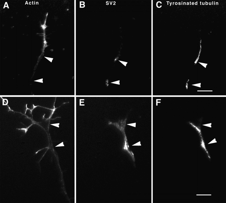
To investigate the role that myosin Va plays in axonal transport of organelles, myosin Va-associated organelle movements were monitored in living neurons using microinjected fluorescently labeled antibodies to myosin Va or expression of a green fluorescent protein-myosin Va tail construct. Myosin Va-associated organelles made rapid bi-directional movements in both normal and dilute-lethal (myosin Va null) neurites. In normal neurons, depolymerization of microtubules by nocodazole slowed, but did not stop movement. In contrast, depolymerization of microtubules in dilute-lethal neurons stopped movement. Myosin Va or synaptic vesicle protein 2 (SV2), which partially colocalizes with myosin Va on organelles, did not accumulate in dilute-lethal neuronal cell bodies because of an anterograde bias associated with organelle transport. However, SV2 showed peripheral accumulations in axon regions of dilute-lethal neurons rich in tyrosinated tubulin. This suggests that myosin Va-associated organelles become stranded in regions rich in dynamic microtubule endings. Consistent with these observations, presynaptic terminals of cerebellar granule cells in dilute-lethal mice showed increased cross-sectional area, and had greater numbers of both synaptic and larger SV2 positive vesicles. Together, these results indicate that myosin Va binds to organelles that are transported in axons along microtubules. This is consistent with both actin- and microtubule-based motors being present on these organelles. Although myosin V activity is not necessary for long-range transport in axons, myosin Va activity is necessary for local movement or processing of organelles in regions, such as presynaptic terminals that lack microtubules.
为了研究肌球蛋白Va在细胞器轴突运输中所起的作用,我们使用微注射的针对肌球蛋白Va的荧光标记抗体或绿色荧光蛋白-肌球蛋白Va尾部构建体的表达,在活神经元中监测了与肌球蛋白Va相关的细胞器运动。在正常和稀致死(肌球蛋白Va缺失)的神经突中,与肌球蛋白Va相关的细胞器都进行快速的双向运动。在正常神经元中,诺考达唑使微管解聚,减慢了但并未停止运动。相比之下,稀致死神经元中的微管解聚则停止了运动。肌球蛋白Va或与肌球蛋白Va在细胞器上部分共定位的突触小泡蛋白2(SV2),由于与细胞器运输相关的顺行偏向,并未在稀致死神经元细胞体中积累。然而,SV2在富含酪氨酸化微管蛋白的稀致死神经元的轴突区域出现外周积累。这表明与肌球蛋白Va相关的细胞器被困在富含动态微管末端的区域。与这些观察结果一致,稀致死小鼠小脑颗粒细胞的突触前终末显示横截面积增加,并且突触小泡和较大的SV2阳性小泡数量更多。总之,这些结果表明肌球蛋白Va与沿微管在轴突中运输的细胞器结合。这与这些细胞器上同时存在基于肌动蛋白和微管的马达是一致的。虽然肌球蛋白V活性对于轴突中的长距离运输不是必需的,但肌球蛋白Va活性对于细胞器在缺乏微管的区域(如突触前终末)的局部运动或加工是必需的。