Wu X, Bowers B, Rao K, Wei Q
Laboratory of Cell Biology, Section on Molecular Cell Biology, National Heart, Lung, and Blood Institute, National Institutes of Health, Bethesda, Maryland 20892, USA.
J Cell Biol. 1998 Dec 28;143(7):1899-918. doi: 10.1083/jcb.143.7.1899.
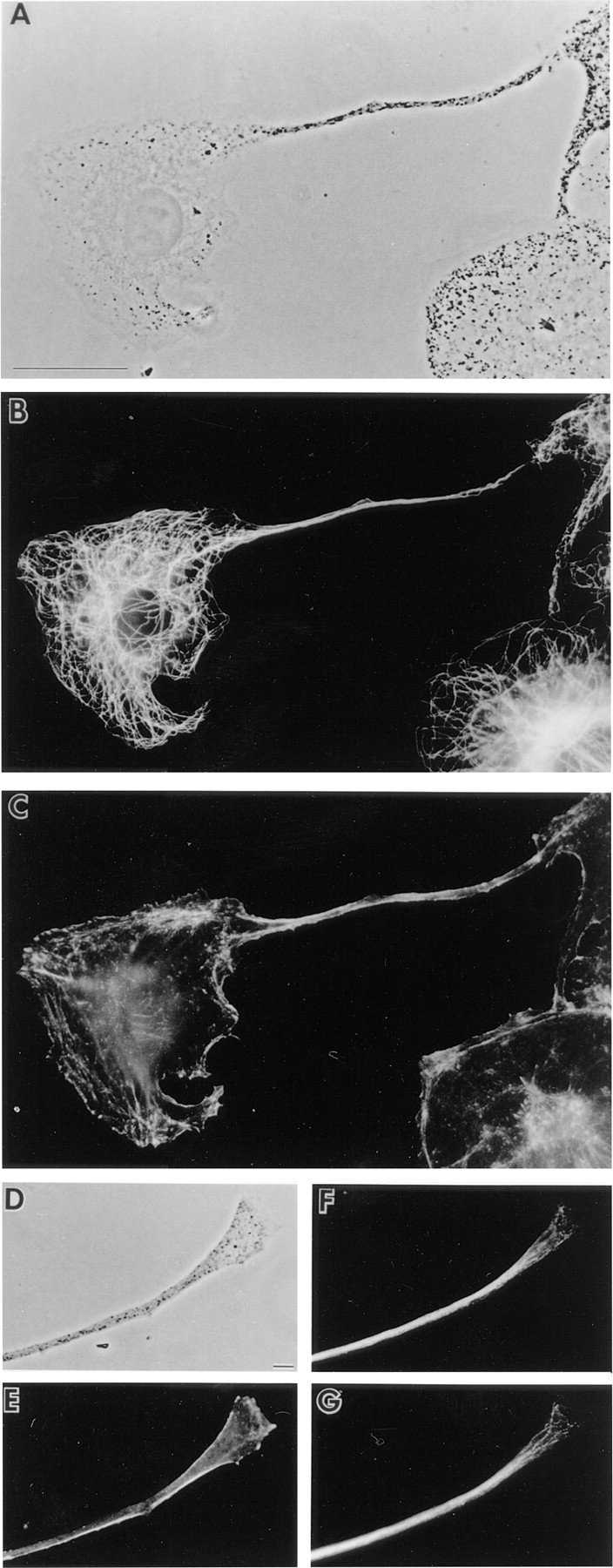
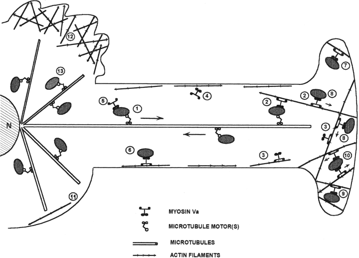
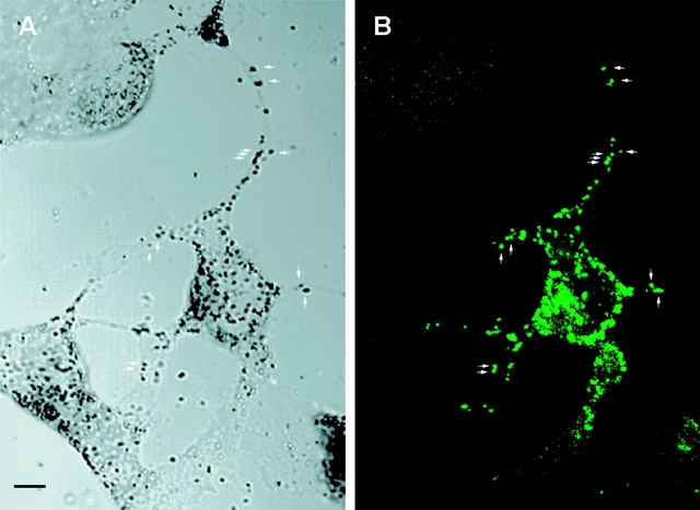
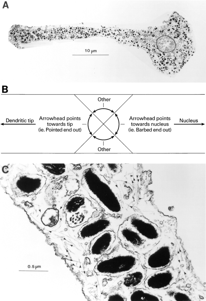
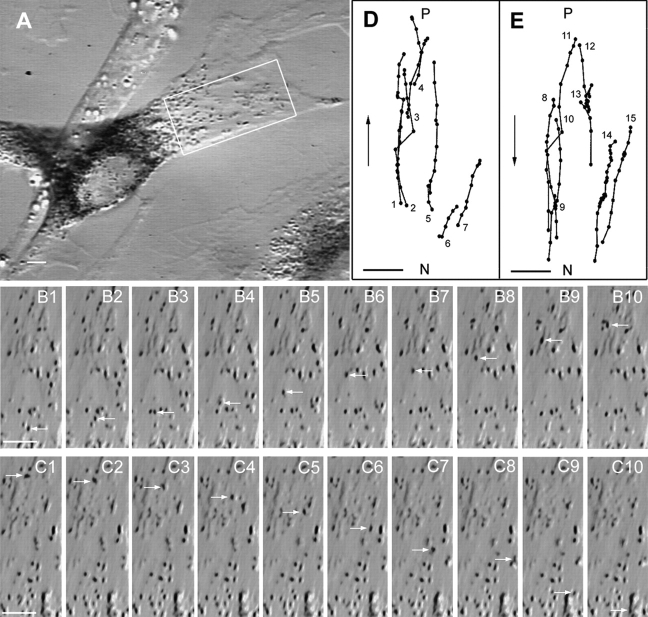
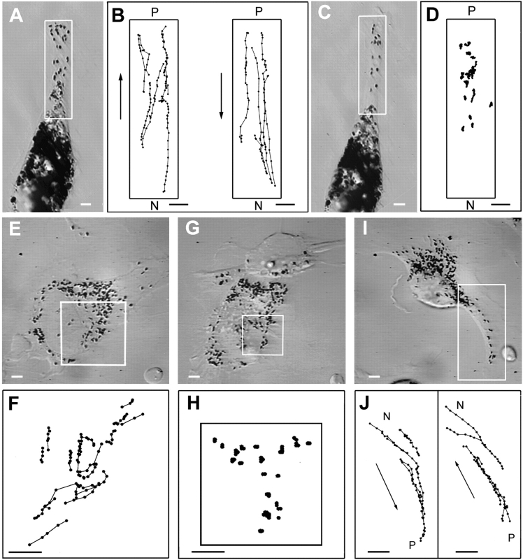
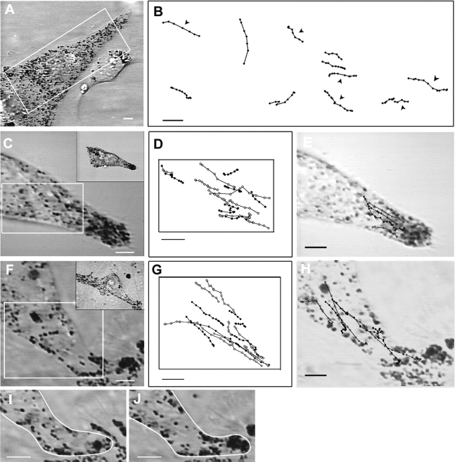
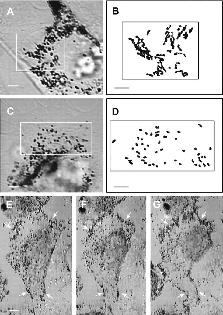
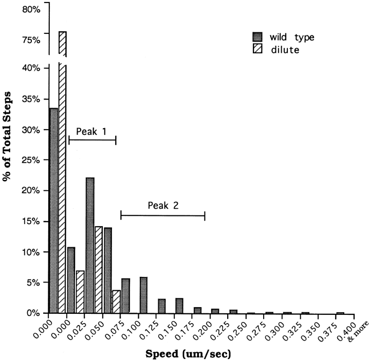
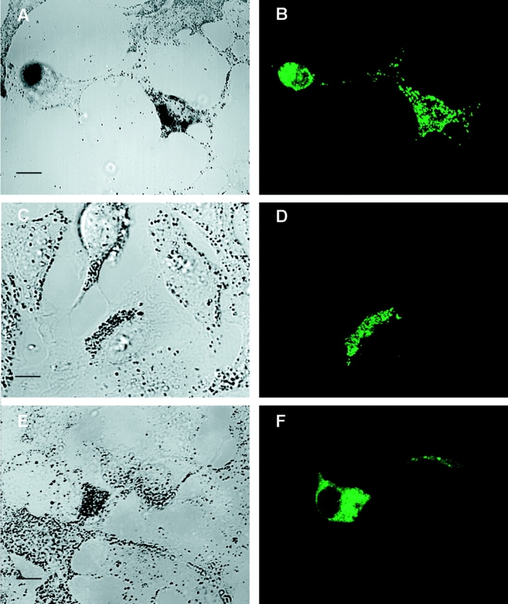
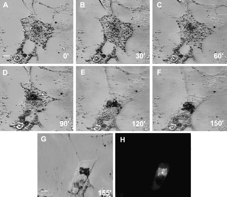
Unlike wild-type mouse melanocytes, where melanosomes are concentrated in dendrites and dendritic tips, melanosomes in dilute (myosin Va-) melanocytes are concentrated in the cell center. Here we sought to define the role that myosin Va plays in melanosome transport and distribution. Actin filaments that comprise a cortical shell running the length of the dendrite were found to exhibit a random orientation, suggesting that myosin Va could drive the outward spreading of melanosomes by catalyzing random walks. In contrast to this mechanism, time lapse video microscopy revealed that melanosomes undergo rapid ( approximately 1.5 microm/s) microtubule-dependent movements to the periphery and back again. This bidirectional traffic occurs in both wild-type and dilute melanocytes, but it is more obvious in dilute melanocytes because the only melanosomes in their periphery are those undergoing this movement. While providing an efficient means to transport melanosomes to the periphery, this component does not by itself result in their net accumulation there. These observations, together with previous studies showing extensive colocalization of myosin Va and melanosomes in the actin-rich periphery, suggest a mechanism in which a myosin Va-dependent interaction of melanosomes with F-actin in the periphery prevents these organelles from returning on microtubules to the cell center, causing their distal accumulation. This "capture" model is supported by the demonstration that (a) expression of the myosin Va tail domain within wild-type cells creates a dilute-like phenotype via a process involving initial colocalization of tail domains with melanosomes in the periphery, followed by an approximately 120-min, microtubule-based redistribution of melanosomes to the cell center; (b) microtubule-dependent melanosome movement appears to be damped by myosin Va; (c) intermittent, microtubule-independent, approximately 0.14 microm/s melanosome movements are seen only in wild-type melanocytes; and (d) these movements do not drive obvious spreading of melanosomes over 90 min. We conclude that long-range, bidirectional, microtubule-dependent melanosome movements, coupled with actomyosin Va-dependent capture of melanosomes in the periphery, is the predominant mechanism responsible for the centrifugal transport and peripheral accumulation of melanosomes in mouse melanocytes. This mechanism represents an alternative to straightforward transport models when interpreting other myosin V mutant phenotypes.
与野生型小鼠黑素细胞不同,野生型小鼠黑素细胞中的黑素体集中在树突和树突尖端,而稀释型(肌球蛋白Va缺陷型)黑素细胞中的黑素体则集中在细胞中心。在此,我们试图确定肌球蛋白Va在黑素体运输和分布中所起的作用。我们发现,构成沿树突全长延伸的皮质壳的肌动蛋白丝呈现随机取向,这表明肌球蛋白Va可能通过催化随机游走驱动黑素体向外扩散。与这种机制不同,延时视频显微镜显示黑素体经历快速(约1.5微米/秒)的依赖微管的向周边移动然后再返回。这种双向运输在野生型和稀释型黑素细胞中均会发生,但在稀释型黑素细胞中更为明显,因为其周边仅有的黑素体就是那些正在进行这种移动的黑素体。虽然这提供了一种将黑素体运输到周边的有效方式,但这一过程本身并不会导致黑素体在那里的净积累。这些观察结果,连同先前的研究表明肌球蛋白Va与黑素体在富含肌动蛋白的周边广泛共定位,提示了一种机制,即黑素体与周边F-肌动蛋白之间的肌球蛋白Va依赖性相互作用可防止这些细胞器通过微管返回细胞中心,从而导致它们在远端积累。这种“捕获”模型得到了以下证据的支持:(a) 在野生型细胞中表达肌球蛋白Va尾域会通过一个过程产生类似稀释型的表型,该过程包括尾域最初与周边黑素体共定位,随后黑素体通过基于微管的方式重新分布到细胞中心,历时约120分钟;(b) 肌球蛋白Va似乎会抑制依赖微管的黑素体移动;(c) 仅在野生型黑素细胞中观察到间歇性的、不依赖微管的、约0.14微米/秒的黑素体移动;(d) 这些移动在90分钟内不会驱动黑素体明显扩散。我们得出结论,长距离、双向、依赖微管的黑素体移动,加上肌动球蛋白Va依赖性的黑素体在周边的捕获,是小鼠黑素细胞中黑素体离心运输和周边积累的主要机制。在解释其他肌球蛋白V突变体表型时,这种机制代表了一种不同于直接运输模型的替代方案。