Iwasaki A, Kelsall B L
Immune Cell Interaction Unit, Mucosal Immunity Section, Laboratory of Clinical Investigation, National Institute of Allergy and Infectious Diseases, National Institutes of Health, Bethesda, Maryland 20892-1890, USA.
J Exp Med. 2000 Apr 17;191(8):1381-94. doi: 10.1084/jem.191.8.1381.
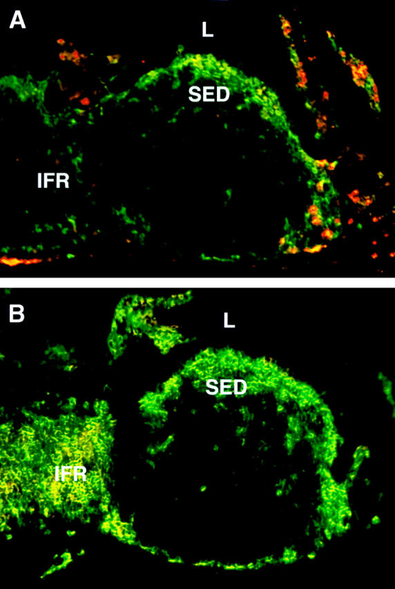
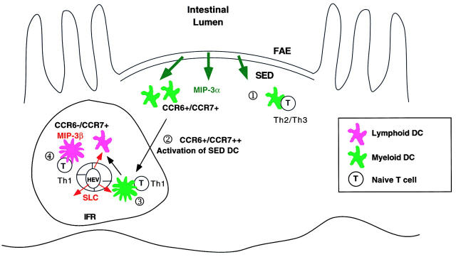
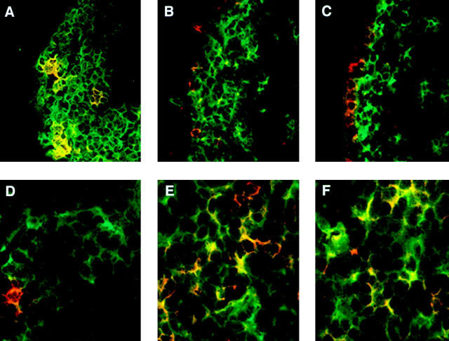
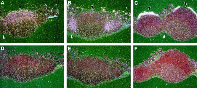
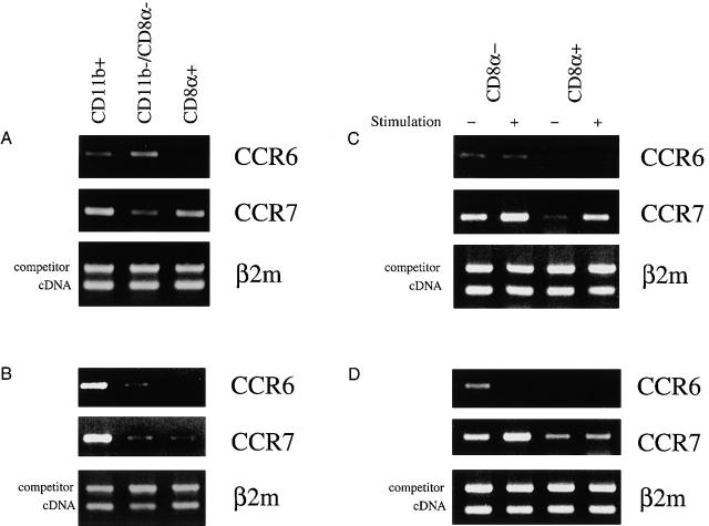
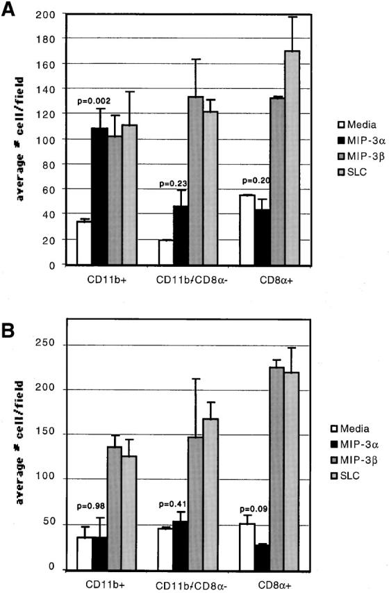
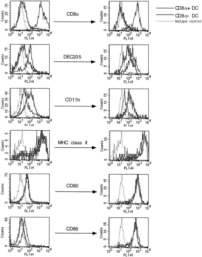
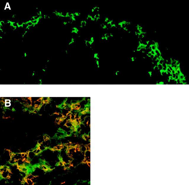
We describe the anatomical localization of three distinct dendritic cell (DC) subsets in the murine Peyer's patch (PP) and explore the role of chemokines in their recruitment. By two-color in situ immunofluorescence, CD11b(+) myeloid DCs were determined to be present in the subepithelial dome (SED) region, whereas CD8alpha(+) lymphoid DCs are present in the T cell-rich interfollicular region (IFR). DCs that lack expression of CD8alpha or CD11b (double negative) are present in both the SED and IFR. By in situ hybridization, macrophage inflammatory protein (MIP)-3alpha mRNA was dramatically expressed only by the follicle-associated epithelium overlying the SED, while its receptor, CCR6, was concentrated in the SED. In contrast, CCR7 was expressed predominantly in the IFR. Consistent with these findings, reverse transcriptase polymerase chain reaction analysis and in vitro chemotaxis assays using freshly isolated DCs revealed that CCR6 was functionally expressed only by DC subsets present in the SED, while all subsets expressed functional CCR7. Moreover, none of the splenic DC subsets migrated toward MIP-3alpha. These data support a distinct role for MIP-3alpha/CCR6 in recruitment of CD11b(+) DCs toward the mucosal surfaces and for MIP-3beta/CCR7 in attraction of CD8alpha(+) DCs to the T cell regions. Finally, we demonstrated that all DC subsets expressed an immature phenotype when freshly isolated and maintained expression of subset markers upon maturation in vitro. In contrast, CCR7 expression by myeloid PP DCs was enhanced with maturation in vitro. In addition, this subset disappeared from the SED and appeared in the IFR after microbial stimulation in vivo, suggesting that immature myeloid SED DCs capture antigens and migrate to IFR to initiate T cell responses after mucosal microbial infections.
我们描述了小鼠派尔集合淋巴结(PP)中三种不同树突状细胞(DC)亚群的解剖定位,并探讨趋化因子在其募集中的作用。通过双色原位免疫荧光法,确定CD11b(+)髓样DC存在于上皮下圆顶(SED)区域,而CD8α(+)淋巴样DC存在于富含T细胞的滤泡间区域(IFR)。缺乏CD8α或CD11b表达的DC(双阴性)同时存在于SED和IFR中。通过原位杂交,巨噬细胞炎性蛋白(MIP)-3α mRNA仅在上覆SED的滤泡相关上皮中显著表达,而其受体CCR6则集中在SED中。相反,CCR7主要在IFR中表达。与这些发现一致,使用新鲜分离的DC进行的逆转录聚合酶链反应分析和体外趋化试验表明,CCR6仅在SED中存在的DC亚群中功能性表达,而所有亚群均表达功能性CCR7。此外,脾脏DC亚群均不向MIP-3α迁移。这些数据支持MIP-3α/CCR6在将CD11b(+) DC募集到黏膜表面中起独特作用,以及MIP-3β/CCR7在将CD8α(+) DC吸引到T细胞区域中起独特作用。最后,我们证明所有DC亚群在新鲜分离时均表现出未成熟表型,并在体外成熟时维持亚群标志物的表达。相比之下,髓样PP DC的CCR7表达在体外成熟时增强。此外,该亚群在体内受到微生物刺激后从SED消失并出现在IFR中,这表明未成熟的髓样SED DC捕获抗原并迁移到IFR以在黏膜微生物感染后启动T细胞反应。