Jin S L, Lee H P, Kim J I, Chin J Y, Choi S J, Joo M, Yum H K
Department of Internal Medicine, Inje University, College of Medicine, Seoul Paik Hospital, Korea.
Korean J Intern Med. 2000 Dec;15(3):240-4. doi: 10.3904/kjim.2000.15.3.240.
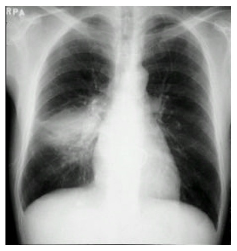
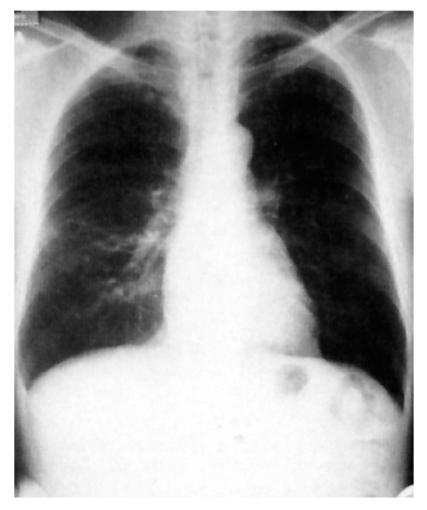
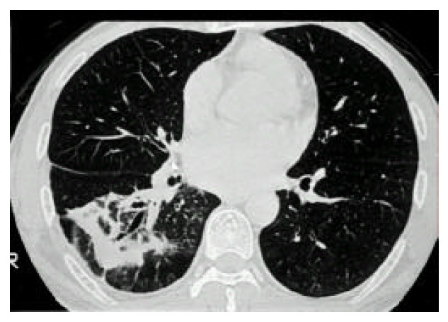
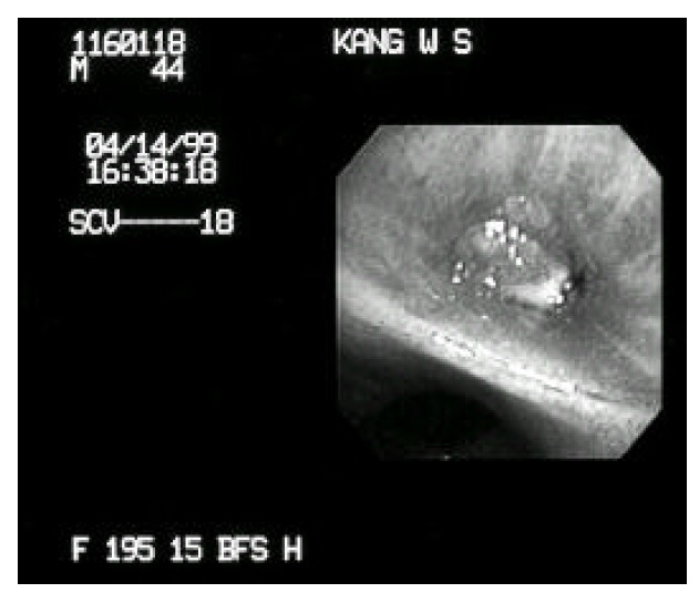
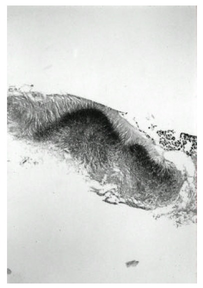
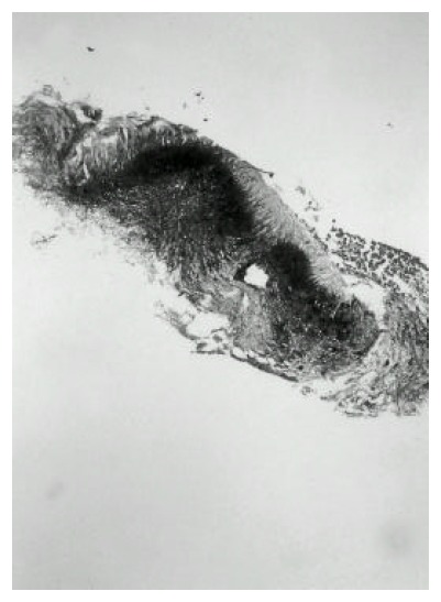
Actinomycosis is an infectious disease caused by certain Actinomyces species. Actinomyces are Gram-positive, non-spore forming organisms characterized by obligate or facultative anaerobic rods that normally inhabit anaerobic niches of the human oral cavity. Cervicofacial, abdominal, pelvic and thoracic infections of Actinomyces are not uncommon, but endobronchial actinomycosis is rarely reported. Endobronchial actinomycosis can be misdiagnosed as unresolving pneumonia, endobronchial lipoma or malignancies. Endobronchial actinomycosis should be included in the differential diagnosis of any endobronchial mass. We report a case of a 43-year-old man who presented with a productive cough and pulmonary consolidation at the right lower lobe on chest radiograph. Fiberoptic bronchoscopy revealed obstruction of the right superior segment of the lower bronchus with an exophytic endobronchial mass. Endobronchial actinomycosis was confirmed by demonstration of sulfur granules in the bronchoscopic biopsy of the mass. Intravenous administration of penicillin G followed by oral amoxacillin/clavulanic acid therapy for 3 months resulted in improving symptoms. Infiltrative consolidation on the chest X-ray was markedly decreased.
放线菌病是一种由某些放线菌属引起的传染病。放线菌是革兰氏阳性、不形成芽孢的微生物,其特征是专性或兼性厌氧杆菌,通常栖息于人类口腔的厌氧环境中。放线菌引起的颈面部、腹部、盆腔和胸部感染并不少见,但支气管内放线菌病很少有报道。支气管内放线菌病可能被误诊为持续性肺炎、支气管内脂肪瘤或恶性肿瘤。任何支气管内肿块的鉴别诊断都应包括支气管内放线菌病。我们报告一例43岁男性患者,其表现为咳痰,胸部X线片显示右下叶肺部实变。纤维支气管镜检查发现下叶支气管右上段有外生性支气管内肿块阻塞。通过在肿块的支气管镜活检中发现硫磺颗粒确诊为支气管内放线菌病。静脉注射青霉素G,随后口服阿莫西林/克拉维酸治疗3个月后症状改善。胸部X线片上的浸润性实变明显减轻。