Presidente P J, McCraw B M, Lumsden J H
Can J Comp Med. 1975 Apr;39(2):166-77.
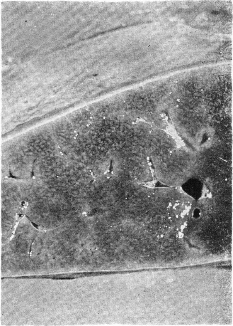
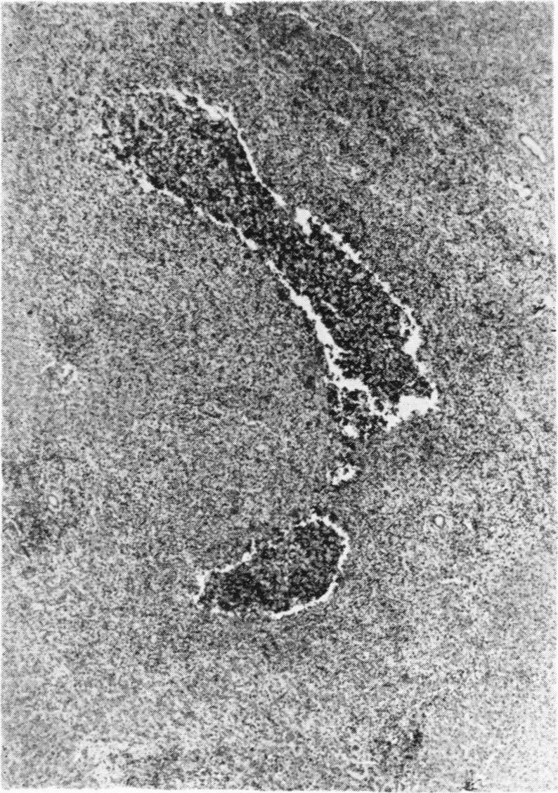
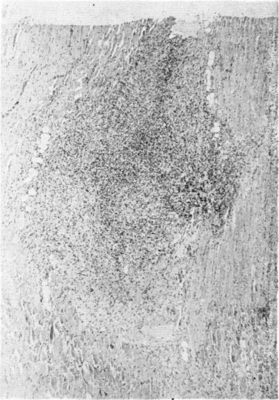
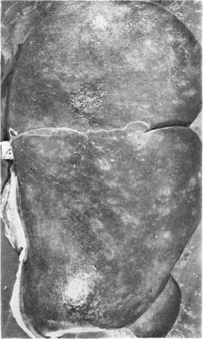
Six white-tailed deer (Odocoileus virginianus) and six sheep were inoculated with metacercariae of Fasciola hepatica. Two animals of each species were given 100, 500 or 2500 metacercariae. One animal in each inocluated group was killed and examined at six weeks postinoculation and the remainder at 15 weeks postinoculation. At six weeks postinoculation the parietal surface of the livers from inoculated deer was covered with gray fibrous plaques and rust colored patches. Fibroplasia with mononuclear cell infiltration characterized Glisson's capsule on the parietal surface. Granulomas were found in the hepatic parenchyma and on the dorsal surface of the lung. Fresh and healing tracks were occasionally found in the liver. In the sheep fibrinous exudate and numerous subcapsular tracks were found on both surfaces of the liver. Inflammatory changes in portal areas and numerous fresh and healing tracks in the hepatic parenchyma were prominent features. At 15 weeks postinoculation inflammatory changes in Glisson's capsule of inoculated deer were less marked than at six weeks but portal fibrosis and hyperplasia of bile duct epithelium were more advanced. A zone of hemorrhage surrounded ducts that contained mature F. hepatica in one deer. The livers from the sheep were rough, pitted and covered with fibrous tags and adhesions to the diaphragm and greater omentum were common. Hemorrhagic tracks were common in the sheep given 500 and 2500 metacercariae. Portal fibrosis and hyperplasia of bile duct epithelium were seen in the sheep (100 metacercariae) that harbored mature F. hepatica.
给六只白尾鹿(弗吉尼亚鹿)和六只绵羊接种肝片吸虫的囊蚴。每种动物中有两只分别接种100、500或2500个囊蚴。每个接种组中的一只动物在接种后六周处死并进行检查,其余的在接种后15周处死并检查。接种后六周,接种鹿的肝脏脏面覆盖着灰色纤维斑块和锈色斑点。脏面的Glisson囊表现为伴有单核细胞浸润的纤维组织增生。在肝实质和肺脏面发现肉芽肿。肝脏中偶尔可见新鲜的和正在愈合的虫道。在绵羊中,肝脏两面均发现有纤维蛋白渗出物和大量被膜下虫道。门管区的炎症变化以及肝实质中大量新鲜的和正在愈合的虫道是突出特征。接种后15周,接种鹿的Glisson囊的炎症变化不如六周时明显,但门管纤维化和胆管上皮增生更明显。在一只鹿中,含有成熟肝片吸虫的胆管周围有一个出血带。绵羊的肝脏粗糙、有凹痕,表面覆盖着纤维条带,与膈肌和大网膜粘连很常见。接种500和2500个囊蚴的绵羊中出血性虫道很常见。在携带成熟肝片吸虫的绵羊(接种100个囊蚴)中可见门管纤维化和胆管上皮增生。