Laukaitis C M, Webb D J, Donais K, Horwitz A F
Department of Cell and Structural Biology, University of Illinois at Urbana-Champaign, Urbana, Illinois, 61801, USA.
J Cell Biol. 2001 Jun 25;153(7):1427-40. doi: 10.1083/jcb.153.7.1427.
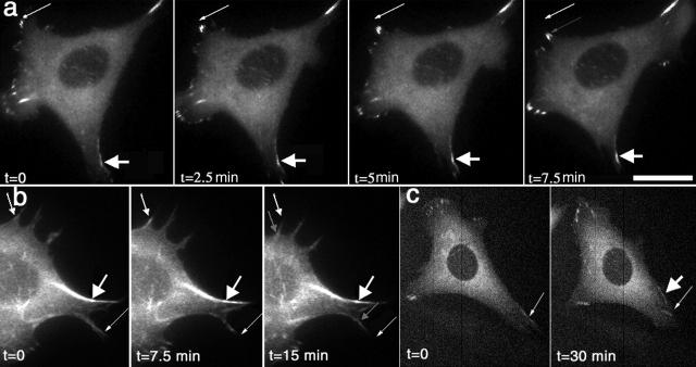
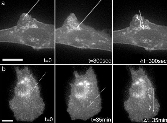
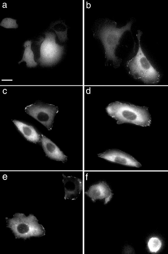
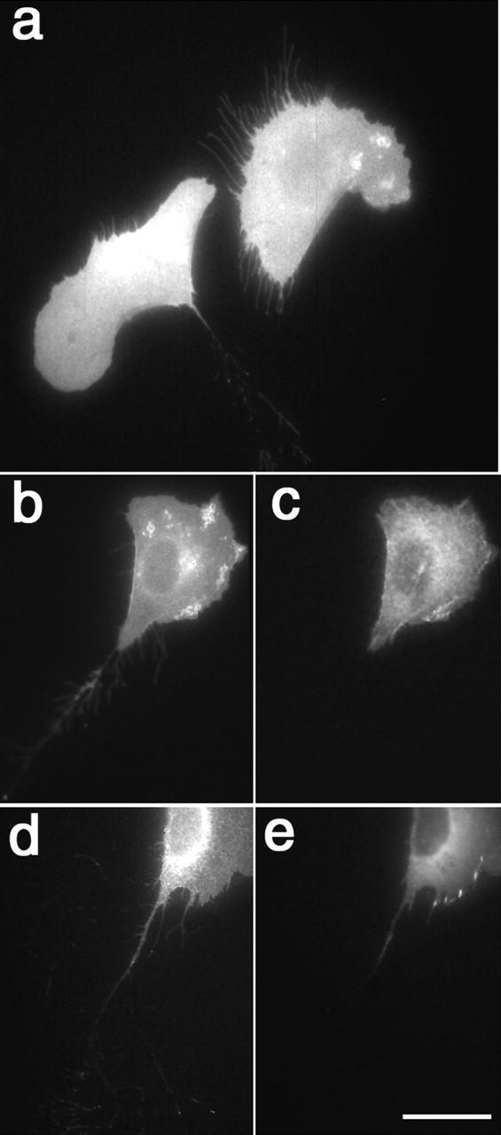
To investigate the mechanisms by which adhesions form and disperse in migrating cells, we expressed alpha 5 integrin, alpha-actinin, and paxillin as green fluorescent protein (GFP) fusions. All localized with their endogenous counterparts and did not perturb migration when expressed at moderate levels. alpha 5-GFP also rescued the adhesive defects in CHO B2 cells, which are alpha 5 integrin deficient. In ruffling cells, alpha 5-GFP and alpha-actinin--GFP localized prominently at the leading edge in membrane protrusions. Of the three GFP fusion proteins that we examined, paxillin was the first component to appear visibly organized in protrusive regions of the cell. When a new protrusion formed, the paxillin appeared to remodel from older to newer adhesions at the leading edge. alpha-Actinin subsequently entered adhesions, which translocated toward the cell center, and inhibited paxillin turnover. The new adhesions formed from small foci of alpha-actinin--GFP and paxillin-GFP, which grew in size. Subsequently, alpha 5 integrin entered the adhesions to form visible complexes, which served to stabilize the adhesions. alpha 5-GFP also resided in endocytic vesicles that emanated from the leading edge of protrusions. Integrin vesicles at the cell rear moved toward the cell body. As cells migrated, alpha 5 vesicles also moved from a perinuclear region to the base of the lamellipodium. The alpha 5 vesicles colocalized with transferrin receptor and FM 4-64 dye. After adhesions broke down in the rear, alpha 5-GFP was found in fibrous structures behind the cell, whereas alpha-actinin--GFP and paxillin-GFP moved up the lateral edge of retracting cells as organized structures and then dissipated.
为了研究黏附在迁移细胞中形成和分散的机制,我们将α5整合素、α-辅肌动蛋白和桩蛋白表达为绿色荧光蛋白(GFP)融合蛋白。所有这些融合蛋白都与它们的内源性对应物共定位,并且在适度表达时不会干扰细胞迁移。α5-GFP还挽救了CHO B2细胞中的黏附缺陷,CHO B2细胞是α5整合素缺陷型细胞。在有皱褶的细胞中,α5-GFP和α-辅肌动蛋白-GFP显著定位于膜突出的前沿。在我们检测的三种GFP融合蛋白中,桩蛋白是第一个在细胞突出区域明显呈现有序排列的成分。当形成新的突出时,桩蛋白似乎在前沿从较旧的黏附重塑为较新的黏附。随后α-辅肌动蛋白进入黏附结构,这些黏附结构向细胞中心移位,并抑制桩蛋白的周转。由α-辅肌动蛋白-GFP和桩蛋白-GFP的小焦点形成新的黏附,其尺寸不断增大。随后,α5整合素进入黏附结构以形成可见的复合物,从而稳定黏附。α5-GFP也存在于从突出前沿发出的内吞小泡中。细胞后部的整合素小泡向细胞体移动。随着细胞迁移,α5小泡也从核周区域移动到片足基部。α5小泡与转铁蛋白受体和FM 4-64染料共定位。在细胞后部的黏附结构解体后,在细胞后方的纤维状结构中发现了α5-GFP,而α-辅肌动蛋白-GFP和桩蛋白-GFP作为有序结构沿着收缩细胞的侧缘向上移动,然后消散。