Mark Karen S, Davis Thomas P
Department of Pharmacology, University of Arizona College of Medicine, Tucson, Arizona 85724-5050, USA.
Am J Physiol Heart Circ Physiol. 2002 Apr;282(4):H1485-94. doi: 10.1152/ajpheart.00645.2001.
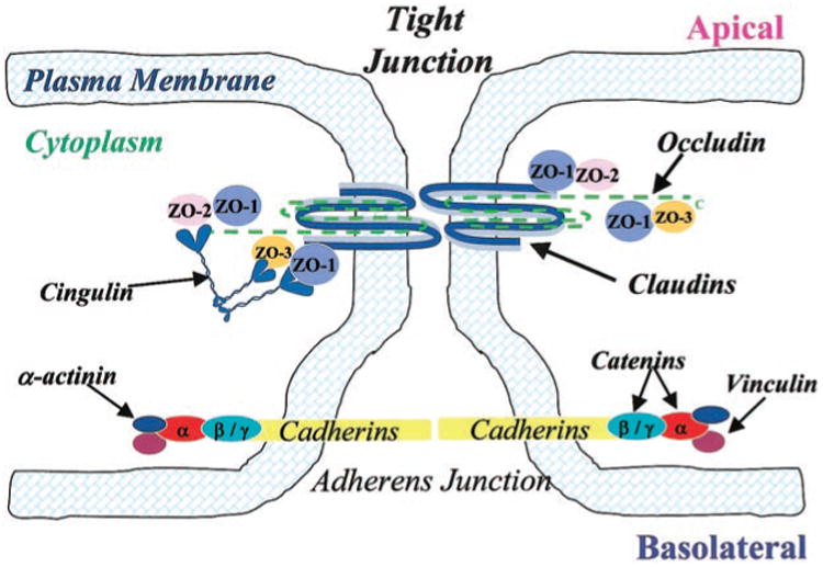
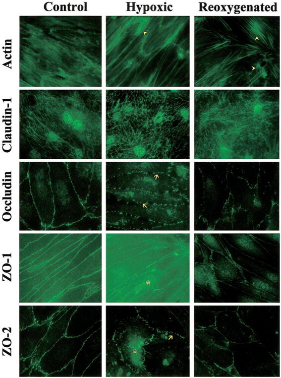
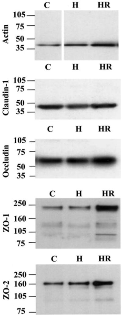
Cerebral microvessel endothelial cells that form the blood-brain barrier (BBB) have tight junctions (TJ) that are critical for maintaining brain homeostasis and low permeability. Both integral (claudin-1 and occludin) and membrane-associated zonula occluden-1 and -2 (ZO-1 and ZO-2) proteins combine to form these TJ complexes that are anchored to the cytoskeletal architecture (actin). Disruptions of the BBB have been attributed to hypoxic conditions that occur with ischemic stroke, pathologies of decreased perfusion, and high-altitude exposure. The effects of hypoxia and posthypoxic reoxygenation in cerebral microvasculature and corresponding cellular mechanisms involved in disrupting the BBB remain unclear. This study examined hypoxia and posthypoxic reoxygenation effects on paracellular permeability and changes in actin and TJ proteins using primary bovine brain microvessel endothelial cells (BBMEC). Hypoxia induced a 2.6-fold increase in [(14)C]sucrose, a marker of paracellular permeability. This effect was significantly reduced (~58%) with posthypoxic reoxygenation. After hypoxia and posthypoxic reoxygenation, actin expression was increased (1.4- and 2.3-fold, respectively). Whereas little change was observed in TJ protein expression immediately after hypoxia, a twofold increase in expression was seen with posthypoxic reoxygenation. Furthermore, immunofluorescence studies showed alterations in occludin, ZO-1, and ZO-2 protein localization during hypoxia and posthypoxic reoxygenation that correlate with the observed changes in BBMEC permeability. The results of this study show hypoxia-induced changes in paracellular permeability may be due to perturbation of TJ complexes and that posthypoxic reoxygenation reverses these effects.
形成血脑屏障(BBB)的脑微血管内皮细胞具有紧密连接(TJ),这对于维持脑内环境稳定和低通透性至关重要。整合蛋白(claudin-1和occludin)以及膜相关的闭合蛋白1和2(ZO-1和ZO-2)共同形成这些紧密连接复合物,并锚定在细胞骨架结构(肌动蛋白)上。血脑屏障的破坏归因于缺血性中风时出现的缺氧状态、灌注减少的病理情况以及高原暴露。缺氧和缺氧后复氧对脑微血管系统的影响以及破坏血脑屏障所涉及的相应细胞机制仍不清楚。本研究使用原代牛脑微血管内皮细胞(BBMEC),研究了缺氧和缺氧后复氧对细胞旁通透性以及肌动蛋白和紧密连接蛋白变化的影响。缺氧使作为细胞旁通透性标志物的[(14)C]蔗糖增加了2.6倍。缺氧后复氧使这种效应显著降低(约58%)。缺氧和缺氧后复氧后,肌动蛋白表达增加(分别增加1.4倍和2.3倍)。缺氧后立即观察到紧密连接蛋白表达变化不大,但缺氧后复氧时表达增加了两倍。此外,免疫荧光研究表明,在缺氧和缺氧后复氧期间,occludin、ZO-1和ZO-2蛋白定位发生改变,这与观察到的BBMEC通透性变化相关。本研究结果表明,缺氧诱导的细胞旁通透性变化可能是由于紧密连接复合物的扰动,而缺氧后复氧可逆转这些效应。