Shen Hong, Zhang Manna, Minuk Gerald Y, Gong Yuewen
Department of Internal Medicine, Faculty of Medicine, University of Manitoba, Winnipeg, Canada.
BMC Cell Biol. 2002 Apr 8;3:9. doi: 10.1186/1471-2121-3-9.
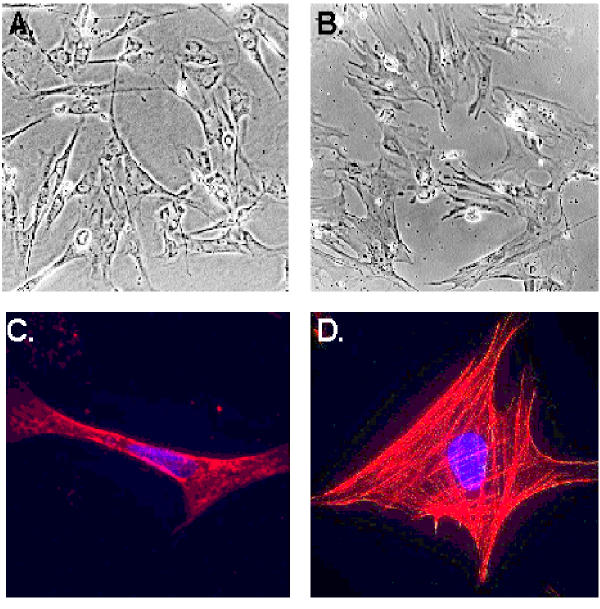
Liver fibrosis is the common sequel of chronic liver diseases. Recent studies have identified hepatic stellate cells as the primary cell type mediating hepatic fibrogenesis. It has been demonstrated that hepatic stellate cells undergo a process of activation during the development of liver fibrosis. During the activation process, hepatic stellate cells acquire myofibroblast-like phenotype featuring the expression of smooth muscle alpha actin. Interferons have been employed for the treatment of viral hepatitis. However, it is unclear what is the effect of interferons on the prevention and treatment of liver fibrosis. Moreover, it is not clear whether there are any differences among interferon alpha, interferon beta, and interferon gamma in the treatment of liver fibrosis. Therefore, our objective in current study is to investigate the effects of rat interferon-alpha, interferon-beta, and interferon-gamma on the proliferation and activation of rat hepatic stellate cells.
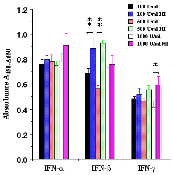
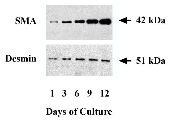
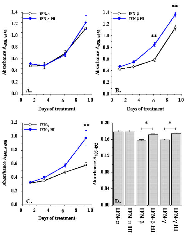
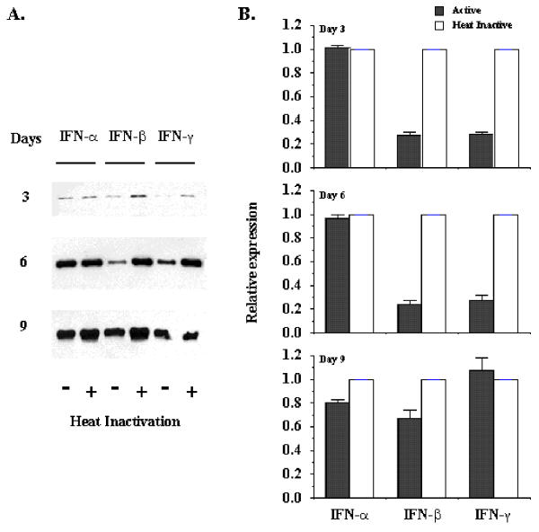
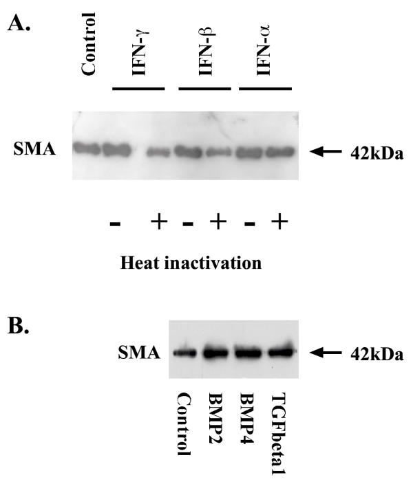
Rat interferon-beta and interferon-gamma significantly inhibited rat hepatic stellate cell proliferation while rat interferon-alpha did not affect the cell proliferation under the same culture condition. Inhibition of cell proliferation was confirmed by both WST-1 cell proliferation assay and 5-bromo-2'-deoxy-uridine incorporation assay. Similar results were observed regarding interferons regulation of hepatic stellate cell activation. Both rat interferon-beta and interferon-gamma reduced smooth muscle alpha-actin abundance after 6 days treatment, but rat interferon-alpha did not alter smooth muscle alpha-actin level.
Our results indicate that rat interferon-alpha and interferon-beta have different biological effects on rat hepatic stellate cells and suggest that there are different signaling events between interferon-alpha and interferon-beta in hepatic stellate cells.
肝纤维化是慢性肝病的常见后遗症。最近的研究已确定肝星状细胞是介导肝纤维化形成的主要细胞类型。已经证明,在肝纤维化发展过程中,肝星状细胞经历激活过程。在激活过程中,肝星状细胞获得具有平滑肌α肌动蛋白表达特征的肌成纤维细胞样表型。干扰素已被用于治疗病毒性肝炎。然而,尚不清楚干扰素对肝纤维化的预防和治疗有何作用。此外,在肝纤维化治疗中,α干扰素、β干扰素和γ干扰素之间是否存在差异也不清楚。因此,我们当前研究的目的是探讨大鼠α干扰素、β干扰素和γ干扰素对大鼠肝星状细胞增殖和激活的影响。
在相同培养条件下,大鼠β干扰素和γ干扰素显著抑制大鼠肝星状细胞增殖,而大鼠α干扰素对细胞增殖无影响。WST-1细胞增殖试验和5-溴-2'-脱氧尿苷掺入试验均证实了细胞增殖受到抑制。关于干扰素对肝星状细胞激活的调节,也观察到了类似结果。处理6天后,大鼠β干扰素和γ干扰素均降低了平滑肌α肌动蛋白丰度,但大鼠α干扰素未改变平滑肌α肌动蛋白水平。
我们的结果表明,大鼠α干扰素和β干扰素对大鼠肝星状细胞具有不同的生物学效应,并提示在肝星状细胞中,α干扰素和β干扰素之间存在不同的信号转导事件。