• 文献检索
  • 文档翻译
  • 深度研究
  • 学术资讯
  • Suppr Zotero 插件Zotero 插件
  • 邀请有礼
  • 套餐&价格
  • 历史记录
应用&插件
Suppr Zotero 插件Zotero 插件浏览器插件Mac 客户端Windows 客户端微信小程序
定价
高级版会员购买积分包购买API积分包
服务
文献检索文档翻译深度研究API 文档MCP 服务
关于我们
关于 Suppr公司介绍联系我们用户协议隐私条款
关注我们

Suppr 超能文献

核心技术专利:CN118964589B侵权必究
粤ICP备2023148730 号-1Suppr @ 2026

文献检索

告别复杂PubMed语法,用中文像聊天一样搜索,搜遍4000万医学文献。AI智能推荐,让科研检索更轻松。

立即免费搜索

文件翻译

保留排版,准确专业,支持PDF/Word/PPT等文件格式,支持 12+语言互译。

免费翻译文档

深度研究

AI帮你快速写综述,25分钟生成高质量综述,智能提取关键信息,辅助科研写作。

立即免费体验

人类手指肌腱的发育

The development of human digital tendons.

作者信息

Chaplin D M, Greenlee T K

出版信息

J Anat. 1975 Nov;120(Pt 2):253-74.

PMID:1201962
原文链接:https://pmc.ncbi.nlm.nih.gov/articles/PMC1231969/
Abstract

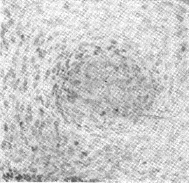
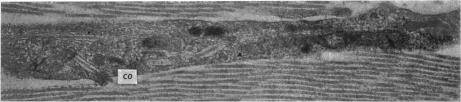
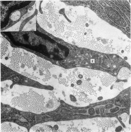
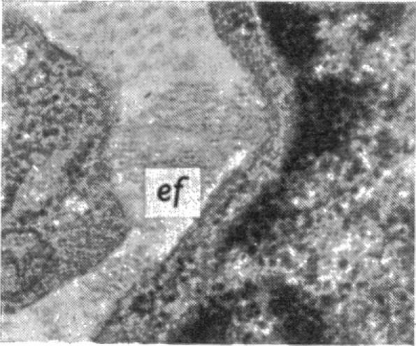
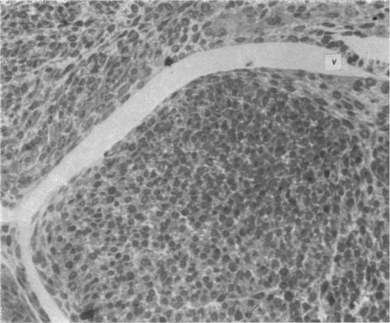
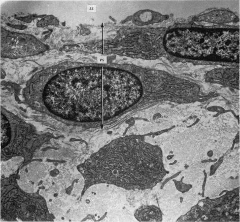
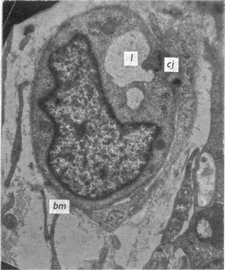
The development of human digital flexor and extensor tendons from 40 days to 112 days of gestation is described. The differentiation of the cell, the formation of collagen fibrils, and their organization into a relatively complex tendon organ are described. This word was supported by a grant from the Graduate Research Fund of the University of Washington. (Materials were obtained from the laboratory for human embryos which is supported by N.I.H. Grant no. HD00836.) We would like to thank Mrs Bea Watts for secretarial assistance.

摘要

本文描述了人类手指屈肌腱和伸肌腱在妊娠40天至112天期间的发育情况。文中阐述了细胞的分化、胶原纤维的形成以及它们如何组织成一个相对复杂的肌腱器官。这项工作得到了华盛顿大学研究生研究基金的资助。(材料取自人类胚胎实验室,该实验室由美国国立卫生研究院资助,资助编号为HD00836。)我们要感谢比阿·瓦茨夫人提供的秘书协助。

https://cdn.ncbi.nlm.nih.gov/pmc/blobs/6e00/1231969/a9c6218a01a7/janat00376-0055-b.jpg
https://cdn.ncbi.nlm.nih.gov/pmc/blobs/6e00/1231969/cf9a8b0a0557/janat00376-0037-a.jpg
https://cdn.ncbi.nlm.nih.gov/pmc/blobs/6e00/1231969/9635d1e5961c/janat00376-0041-b.jpg
https://cdn.ncbi.nlm.nih.gov/pmc/blobs/6e00/1231969/1fd737256312/janat00376-0046-a.jpg
https://cdn.ncbi.nlm.nih.gov/pmc/blobs/6e00/1231969/035ac89ea1cb/janat00376-0046-b.jpg
https://cdn.ncbi.nlm.nih.gov/pmc/blobs/6e00/1231969/4d8e2be25dc1/janat00376-0047-a.jpg
https://cdn.ncbi.nlm.nih.gov/pmc/blobs/6e00/1231969/7e2c054157b8/janat00376-0048-a.jpg
https://cdn.ncbi.nlm.nih.gov/pmc/blobs/6e00/1231969/060c8882badd/janat00376-0049-a.jpg
https://cdn.ncbi.nlm.nih.gov/pmc/blobs/6e00/1231969/e921c6183874/janat00376-0049-b.jpg
https://cdn.ncbi.nlm.nih.gov/pmc/blobs/6e00/1231969/bb4f708fe869/janat00376-0050-a.jpg
https://cdn.ncbi.nlm.nih.gov/pmc/blobs/6e00/1231969/260f6f4d8d14/janat00376-0050-b.jpg
https://cdn.ncbi.nlm.nih.gov/pmc/blobs/6e00/1231969/c6736332ed73/janat00376-0051-a.jpg
https://cdn.ncbi.nlm.nih.gov/pmc/blobs/6e00/1231969/ee006285a373/janat00376-0052-a.jpg
https://cdn.ncbi.nlm.nih.gov/pmc/blobs/6e00/1231969/d5fde62ca2d2/janat00376-0054-a.jpg
https://cdn.ncbi.nlm.nih.gov/pmc/blobs/6e00/1231969/a97a21c60a54/janat00376-0055-a.jpg
https://cdn.ncbi.nlm.nih.gov/pmc/blobs/6e00/1231969/a9c6218a01a7/janat00376-0055-b.jpg

相似文献

1
The development of human digital tendons.人类手指肌腱的发育
J Anat. 1975 Nov;120(Pt 2):253-74.
2
A fine structural study of the development of the chick flexor digital tendon: a model for synovial sheathed tendon healing.鸡趾屈肌腱发育的超微结构研究:滑膜鞘肌腱愈合的模型
Am J Anat. 1975 Jul;143(3):303-13. doi: 10.1002/aja.1001430304.
3
Chick vincula: elastic structures with a check-rein mechanism.鸡的系带:具有止动机制的弹性结构。
J Anat. 1975 Apr;119(Pt 2):295-308.
4
[Fibroblast modulation in the course of tendon regeneration].[肌腱再生过程中的成纤维细胞调节]
Verh Anat Ges. 1977(71 Pt 1):477-83.
5
Fine structure of the lens epithelium.
Ann Ophthalmol. 1975 May;7(5):623-6.
6
Changes of nuclear structure in cells of the developing Xenopus embryo.
Acta Morphol Acad Sci Hung. 1974;22(2):203-11.
7
[Demonstration, by means of electron microscopy, of the penetration of somitic cells into the mesoblast of the limb buds of reptile embryos (Anguis fragilis, Lacerta viridis)].[通过电子显微镜对爬行类胚胎(脆蛇蜥、绿蜥蜴)肢体芽中体节细胞向中胚层的渗透进行的演示]
Arch Anat Microsc Morphol Exp. 1975;64(4):287-316.
8
Ultrastructural differentiation of Leydig cells in the testis of 17-day-old chick embryo and newly hatched chickens.17日龄鸡胚和新孵出雏鸡睾丸中莱迪希细胞的超微结构分化
Prog Clin Biol Res. 1981;59B:363-72.
9
Histology and ultrastructure of the developing superficial digital flexor tendon in rabbits.兔发育过程中浅屈肌腱的组织学与超微结构
Anat Histol Embryol. 2008 Apr;37(2):134-40. doi: 10.1111/j.1439-0264.2007.00811.x.
10
Amphophil adenoma of the human pituitary gland with masses of cytoplasmic microfilaments.伴有大量细胞质微丝的人垂体嗜酸性腺瘤。
Endokrinologie. 1974 Sep;63(3):402-8.

引用本文的文献

1
Subsynovial connective tissue development in the rabbit carpal tunnel.兔腕管滑膜下结缔组织发育。
Vet Med Sci. 2020 Nov;6(4):1025-1033. doi: 10.1002/vms3.281. Epub 2020 May 6.
2
Models of tendon development and injury.肌腱发育与损伤模型。
BMC Biomed Eng. 2019;1. doi: 10.1186/s42490-019-0029-5. Epub 2019 Nov 29.
3
Cell-cell junctions in developing and adult tendons.发育中和成年肌腱中的细胞间连接。

本文引用的文献

1
A modified procedure for lead staining of thin sections.一种用于薄切片铅染色的改良方法。
J Biophys Biochem Cytol. 1961 Dec;11(3):736-9. doi: 10.1083/jcb.11.3.736.
2
BLOOD SUPPLY OF TENDONS.肌腱的血液供应
Am J Surg. 1965 Mar;109:272-6. doi: 10.1016/s0002-9610(65)80073-3.
3
Fibroblasts and collagen.成纤维细胞与胶原蛋白。
Tissue Barriers. 2020;8(1):1695491. doi: 10.1080/21688370.2019.1695491. Epub 2019 Dec 9.
4
Multiple musculovascular anomalies in the superior extremities of a cadaver: a case report.一具尸体上肢的多处肌肉血管异常:病例报告
J Clin Diagn Res. 2013 Feb;7(2):342-6. doi: 10.7860/JCDR/2013/4672.2763. Epub 2013 Feb 1.
5
Preferential cell response to anisotropic electro-spun fibrous scaffolds under tension-free conditions.在无张力条件下,细胞对各向异性静电纺纤维支架的优先响应。
J Mater Sci Mater Med. 2012 Jan;23(1):137-48. doi: 10.1007/s10856-011-4471-8. Epub 2011 Nov 22.
6
Ultrastructural studies on the gastrocnemius tendon of selenium-deficient ducklings.缺硒雏鸭腓肠肌腱的超微结构研究
Am J Pathol. 1981 May;103(2):201-9.
7
Attachment and extracellular matrix differences between tendon and synovial fibroblastic cells.肌腱和成纤维滑膜细胞之间的黏附及细胞外基质差异
In Vitro. 1983 Feb;19(2):127-33. doi: 10.1007/BF02621896.
8
Extracellular compartments in tendon morphogenesis: collagen fibril, bundle, and macroaggregate formation.肌腱形态发生中的细胞外区室:胶原原纤维、束和大聚集体的形成。
J Cell Biol. 1986 Jul;103(1):231-40. doi: 10.1083/jcb.103.1.231.
9
Intracellular collagen fibrils: evidence of an intracellular source from experiments with tendon fibroblasts and fibroblastic tumour cells.细胞内胶原纤维:来自肌腱成纤维细胞和成纤维细胞瘤细胞实验的细胞内来源证据。
J Anat. 1988 Jun;158:1-12.
Br Med Bull. 1962 Sep;18:233-7. doi: 10.1093/oxfordjournals.bmb.a069985.
4
Improvements in epoxy resin embedding methods.环氧树脂包埋方法的改进。
J Biophys Biochem Cytol. 1961 Feb;9(2):409-14. doi: 10.1083/jcb.9.2.409.
5
Embedding in epoxy resins for ultrathin sectioning in electron microscopy.嵌入环氧树脂用于电子显微镜超薄切片。
Stain Technol. 1960 Nov;35:313-23. doi: 10.3109/10520296009114754.
6
The blood supply of the flexor and extensor tendons of the fingers in man.
J Bone Joint Surg Br. 1953 Feb;35-B(1):131-8. doi: 10.1302/0301-620X.35B1.131.
7
The development of the rat flexor digital tendon, a fine structure study.大鼠屈指肌腱的发育:一项精细结构研究
J Ultrastruct Res. 1967 May;18(3):354-76. doi: 10.1016/s0022-5320(67)80124-2.
8
Electron microscopy: attachment sites between connective tissue cells.电子显微镜检查:结缔组织细胞之间的附着位点。
Science. 1966 Aug 26;153(3739):997-9. doi: 10.1126/science.153.3739.997.
9
The vascular anatomy within normal tendons, divided tendons, free tendon grafts and pedicle tendon grafts in rabbits. A microradioangiographic study.
J Bone Joint Surg Br. 1973 May;55(2):369-89.
10
The fibroblast and wound repair.成纤维细胞与伤口修复。
Biol Rev Camb Philos Soc. 1968 Feb;43(1):51-96. doi: 10.1111/j.1469-185x.1968.tb01109.x.