Muse K E, Powell D A, Collier A M
Infect Immun. 1976 Jan;13(1):229-37. doi: 10.1128/iai.13.1.229-237.1976.
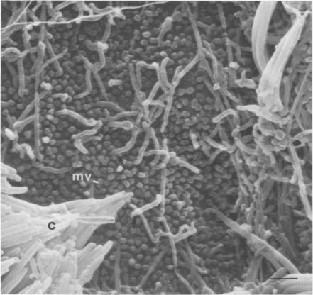
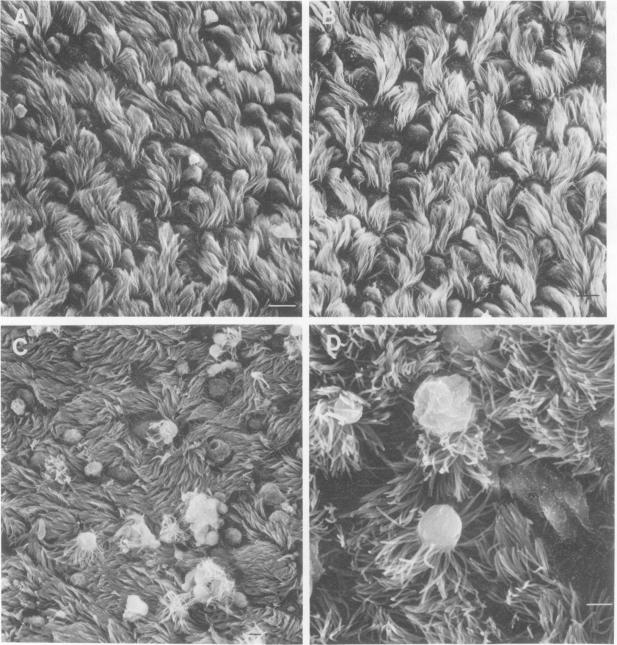
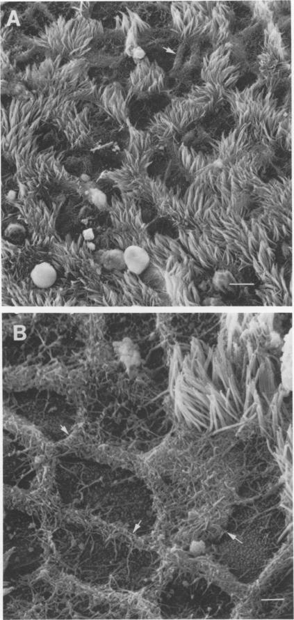
Hamster tracheal rings in organ culture were inoculated with a virulent strain of Mycoplasma pneumoniae and examined by scanning electron microscopy. A progressive increase in epithelial cell injury was detected from 48 to 96 h post-inoculation and was characterized by apparent loss of the apical portion of ciliated cells. M. pneumoniae attaching to the epithelial cell surfaces could be identified by comparison with the surface morphology of mycoplasmas grown on glass cover slips.
将器官培养中的仓鼠气管环接种肺炎支原体强毒株,并通过扫描电子显微镜进行检查。在接种后48至96小时检测到上皮细胞损伤逐渐增加,其特征是纤毛细胞顶端部分明显缺失。通过与生长在玻璃盖玻片上的支原体表面形态进行比较,可以识别附着在上皮细胞表面的肺炎支原体。