Harris J L, Howe H B, Roth I L
J Bacteriol. 1975 Jun;122(3):1239-46. doi: 10.1128/jb.122.3.1239-1246.1975.
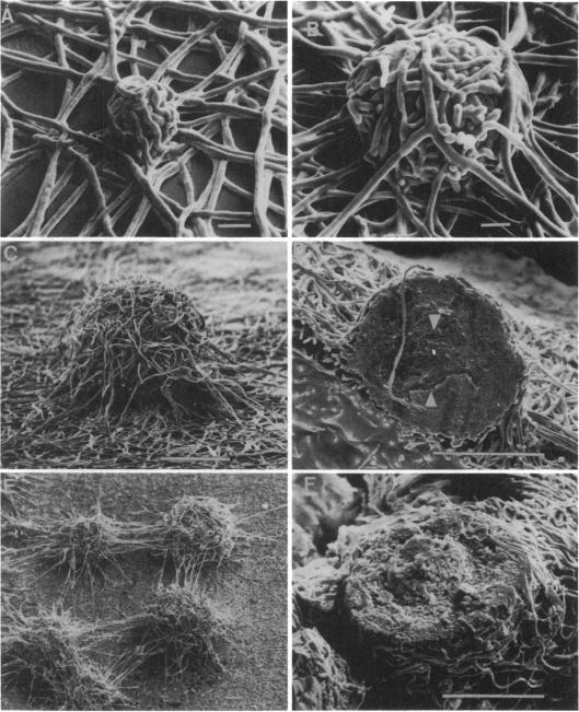
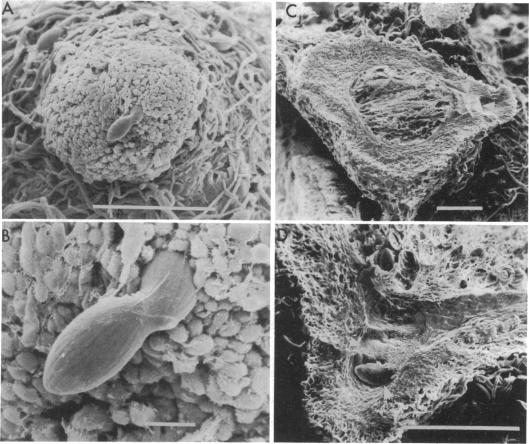
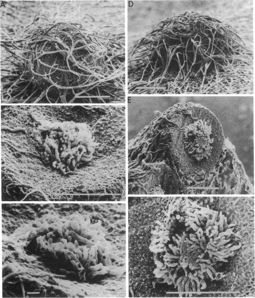
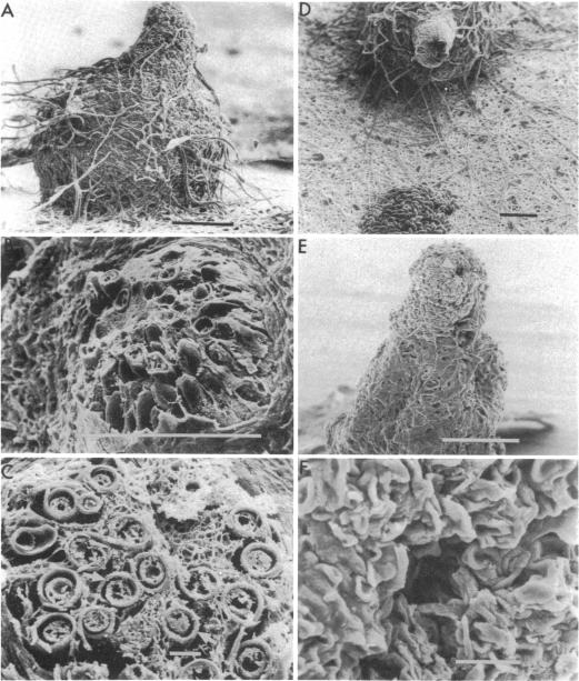
Stages in the development of perithecia of Neurospora crassa, designated by the time elapsed after crossing, were investigated with the scanning electron microscope, from protoperithecia through perithecia. The usual examination of external features of whole specimens with this instrument was augmented by a freeze-fracture technique which allowed the viewing of development internally as well. Rapid increases in perithecial size soon after crossing were followed by the appearance, in section, of a centrum, at first undifferentiated but subsequently developing ascogenous hyphae. The perithecial beak appeared as a compact mass easily distinguishable in whole specimens from the surrounding hyphae by means of texture as well as shape. Two ascospores were photographed during emergence from an ostiole, but ostioles were found more frequently closed than open.
利用扫描电子显微镜对粗糙脉孢菌子囊壳发育阶段(根据杂交后经过的时间来界定)进行了研究,从原雌器到子囊壳。用该仪器对整个标本的外部特征进行常规检查时,采用了冷冻断裂技术,这使得也能从内部观察发育情况。杂交后不久,子囊壳大小迅速增加,随后在切片中出现了子实层,起初未分化,但随后发育出产囊丝。子囊壳喙表现为紧密的一团,通过质地和形状,在整个标本中很容易与周围菌丝区分开来。在两个子囊孢子从子囊口出现时进行了拍照,但发现子囊口关闭的情况比开放的情况更常见。