Bender Roland A, Dubé Celine, Gonzalez-Vega Rebeca, Mina Erene W, Baram Tallie Z
Department of Anatomy and Neurobiology, University of California at Irvine, Irvine, California 92697-4475, USA.
Hippocampus. 2003;13(3):399-412. doi: 10.1002/hipo.10089.
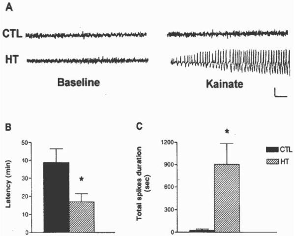
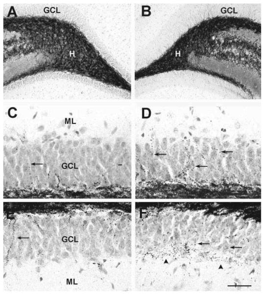
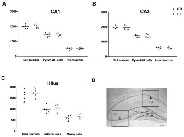
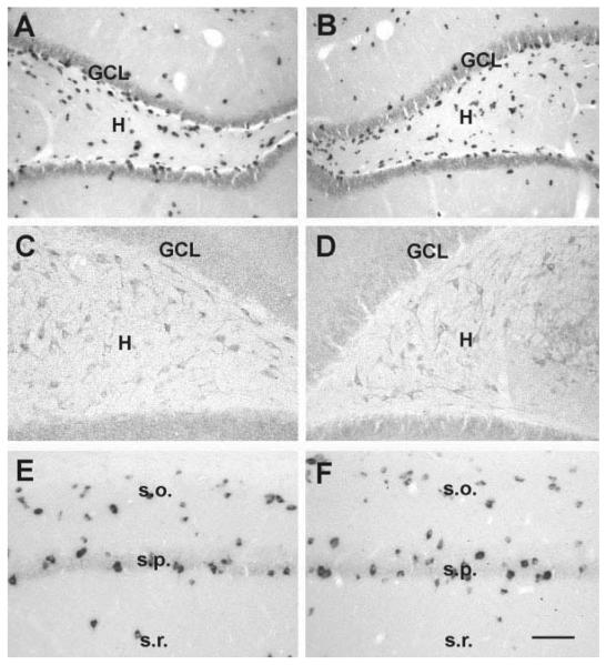
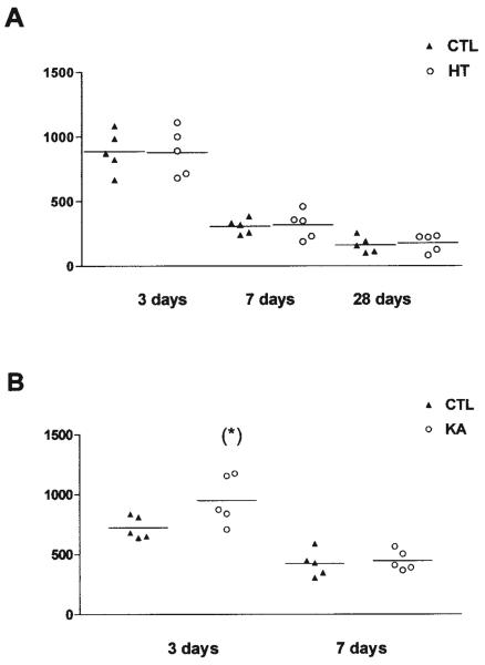
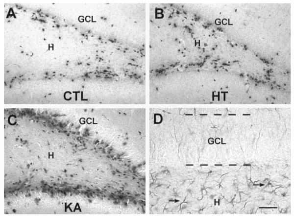
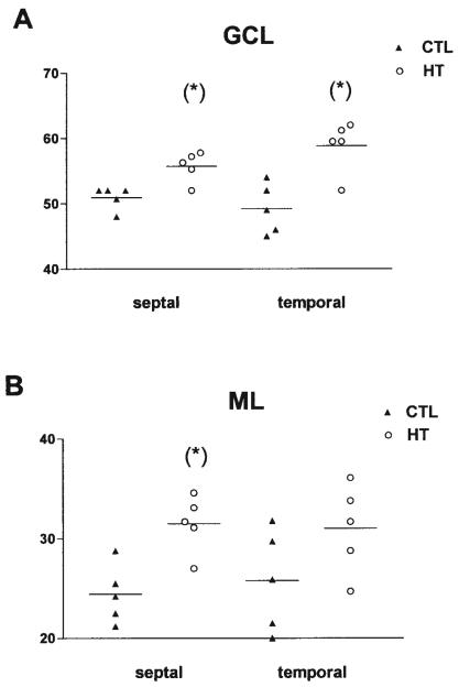
Seizures induced by fever (febrile seizures) are the most frequent seizures affecting infants and children; however, their impact on the developing hippocampal formation is not completely understood. Such understanding is highly important because of the potential relationship of prolonged febrile seizures to temporal lobe epilepsy. Using an immature rat model, we have previously demonstrated that prolonged experimental febrile seizures render the hippocampus hyperexcitable throughout life. Here we examined whether (1) neuronal loss, (2) altered neurogenesis, or (3) mossy fiber sprouting, all implicated in epileptogenesis in both animal models and humans, were involved in the generation of a pro-epileptic, hyperexcitable hippocampus by these seizures. The results demonstrated that prolonged experimental febrile seizures did not result in appreciable loss of any vulnerable hippocampal cell population, though causing strikingly enhanced sensitivity to hippocampal excitants later in life. In addition, experimental febrile seizures on postnatal day 10 did not enhance proliferation of granule cells, whereas seizures generated by kainic acid during the same developmental age increased neurogenesis in the immature hippocampus. However, prolonged febrile seizures resulted in long-term axonal reorganization in the immature hippocampal formation: Mossy fiber densities in granule cell- and molecular layers were significantly increased by 3 months (but not 10 days) after the seizures. Thus, the data indicate that prolonged febrile seizures influence connectivity of the immature hippocampus long-term, and this process requires neither significant neuronal loss nor altered neurogenesis. In addition, the temporal course of the augmented mossy fiber invasion of the granule cell and molecular layers suggests that it is a consequence, rather than the cause, of the hyperexcitable hippocampal network resulting from these seizures.
发热引起的惊厥(热性惊厥)是影响婴幼儿最常见的惊厥类型;然而,其对发育中的海马结构的影响尚未完全明确。鉴于长时间热性惊厥与颞叶癫痫之间可能存在的关联,这种理解至关重要。我们先前利用未成熟大鼠模型证明,长时间的实验性热性惊厥会使海马终生兴奋性增强。在此,我们研究了以下因素是否参与了由这些惊厥导致的促癫痫、兴奋性增强的海马的形成:(1)神经元丢失;(2)神经发生改变;或(3)苔藓纤维出芽,这些因素在动物模型和人类癫痫发生中均有涉及。结果表明,长时间的实验性热性惊厥并未导致任何易损海马细胞群出现明显丢失,尽管在后期会使海马对兴奋性物质的敏感性显著增强。此外,出生后第10天的实验性热性惊厥并未增强颗粒细胞的增殖,而相同发育阶段由 kainic 酸引发的惊厥则增加了未成熟海马中的神经发生。然而,长时间热性惊厥导致未成熟海马结构出现长期轴突重组:惊厥后3个月(而非10天),颗粒细胞层和分子层中的苔藓纤维密度显著增加。因此,数据表明长时间热性惊厥会长期影响未成熟海马的连接性,且这一过程既不需要显著的神经元丢失,也不需要神经发生改变。此外,颗粒细胞层和分子层中苔藓纤维侵入增加的时间进程表明,它是这些惊厥导致的海马兴奋性增强网络的结果,而非原因。