Jang Hyun-Jung, Lim Hyo K, Lee Won Jae, Kim Seong Hyun, Kim Min Ju, Choi Dongil, Lee Soon Jin, Lim Jae Hoon
Department of Radiology and Center for Imaging Science, Samsung Medical Center, Sungkyunkwan University School of Medicine, Kangnam-gu, Seoul, Korea.
Korean J Radiol. 2003 Apr-Jun;4(2):91-100. doi: 10.3348/kjr.2003.4.2.91.
To determine the findings of various focal hepatic lesions at contrast-enhanced gray-scale ultrasound (US) using a coded harmonic angio (CHA) technique and emphasizing lesion characterization.
The study involved 95 patients with 105 focal hepatic lesions, namely 51 hepatocellular carcinomas (HCCs), 22 metastases, 22 hemangiomas, four cases of focal nodular hyperplasia (FNH), and six nontumorous nodules. After the injection of a microbubble contrast agent (SH U 508A), gray-scale harmonic US studies using a CHA technique were performed with a combination of continuous scanning to assess the intratumoral vasculature (vascular imaging) and interval-delay scanning to determine the sequential enhancement pattern (acoustic emission imaging). Each imaging pattern was categorized and analyzed.
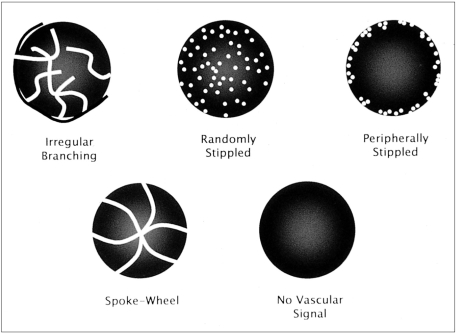
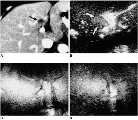
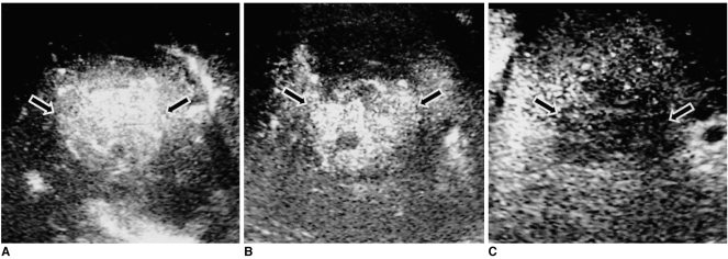
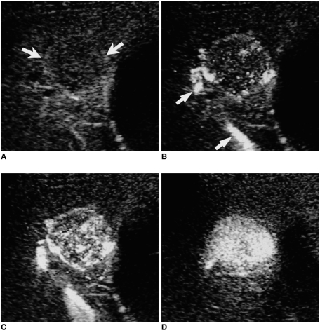
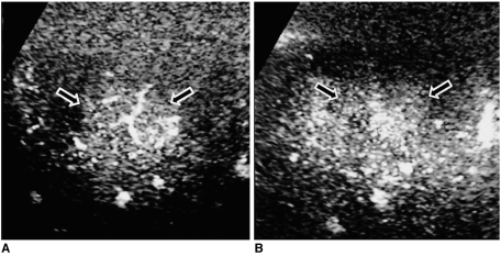
At vascular imaging, 69% of HCCs (35/51) showed irregular branching vessels, while in 91% of metastases (20/22) a peripherally stippled pattern was observed. Intratumoral vessels were absent in 95% of hemangiomas (21/22) and all nontumorous lesions (6/6), while in 75% of FNHs (3/4) a spoke-wheel pattern was evident. At acoustic emission imaging, 71% of HCCs (36/51) showed heterogeneous enhancement and 86% (19/22) of metastases showed rim- or flame-like peripheral enhancement during the early phase, with washout occurring in all HCCs and metastases (100%, 73/73) during the late phase. In hemangiomas, enhancement was either peripheral and nodular (19/22, 86%) or persistent and homogeneous (3/22, 14%), and 75% of FNHs (3/4) became isoechoic during the late phase.
At contrast-enhanced gray-scale US using a CHA technique, a period of continuous scanning depicted the intratumoral vasculature, and interval-delay scanning demonstrated the sequential enhancement pattern. The characteristic findings of various focal hepatic lesions were thus determined.
采用编码谐波血管造影(CHA)技术并着重进行病变特征分析,以确定不同肝脏局灶性病变在灰阶超声造影(US)检查中的表现。
本研究纳入95例患者,共105个肝脏局灶性病变,其中肝细胞癌(HCC)51例、转移瘤22例、肝血管瘤22例、局灶性结节性增生(FNH)4例以及非肿瘤性结节6例。注射微泡造影剂(SH U 508A)后,采用CHA技术进行灰阶谐波超声检查,结合连续扫描评估瘤内血管系统(血管成像)以及间隔延迟扫描以确定序列增强模式(声发射成像)。对每种成像模式进行分类和分析。
在血管成像中,69%的HCC(35/51)显示不规则分支血管,而91%的转移瘤(20/22)观察到周边点状模式。95%的肝血管瘤(21/22)和所有非肿瘤性病变(6/6)瘤内无血管,而75%的FNH(3/4)可见辐轮状模式。在声发射成像中,71%的HCC(36/51)表现为不均匀增强,86%的转移瘤(19/22)在早期表现为边缘或火焰状周边增强,所有HCC和转移瘤(100%,73/73)在晚期均出现廓清。肝血管瘤的增强方式为周边结节状(19/22,86%)或持续均匀增强(3/22,14%),75%的FNH(3/4)在晚期变为等回声。
采用CHA技术的灰阶超声造影检查中,连续扫描可显示瘤内血管系统,间隔延迟扫描可显示序列增强模式。由此确定了不同肝脏局灶性病变的特征性表现。