Choi Dongil, Lim Hyo K, Lee Won Jae, Kim Seung Hoon, Kim Min Ju, Kim Seung Kwon, Jang Kyung Mi, Lee Ji Yeon, Lim Jae Hoon
Department of Radiology, Samsung Medical Center, Sungkyunkwan University School of Medicine, Kangnam-gu, Seoul, Korea.
Korean J Radiol. 2004 Jul-Sep;5(3):185-98. doi: 10.3348/kjr.2004.5.3.185.
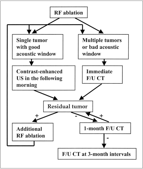
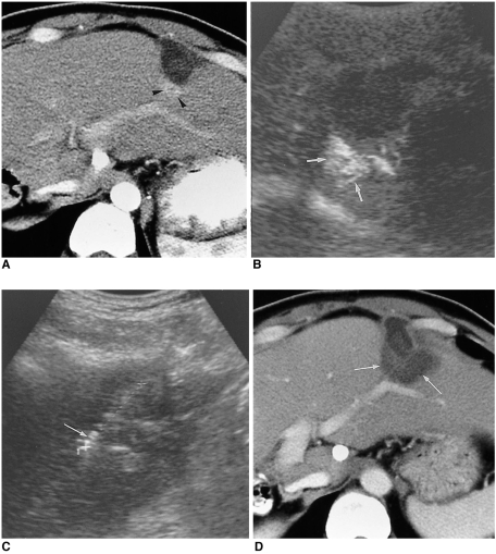
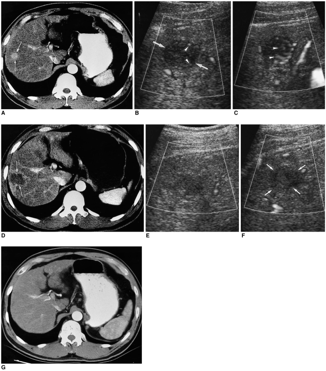
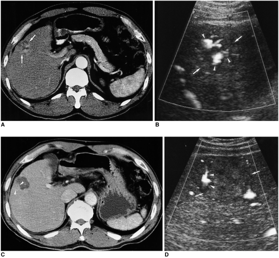
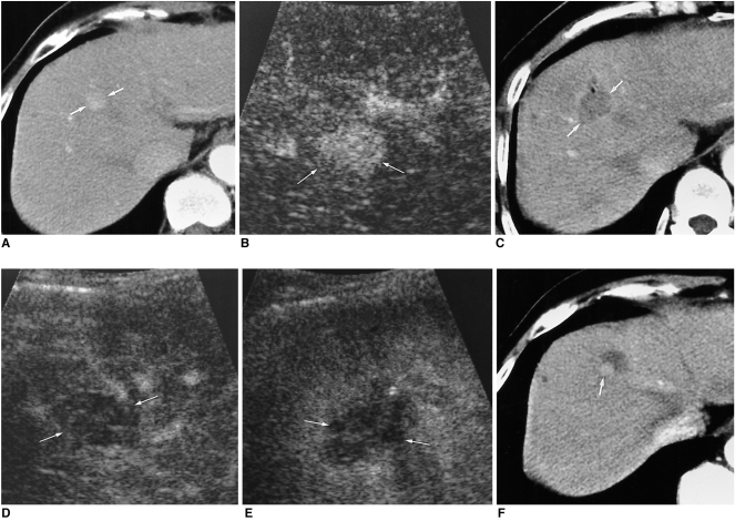
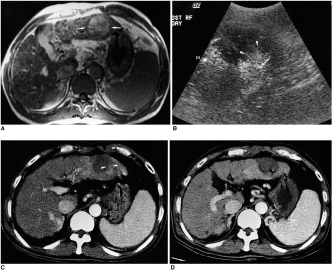
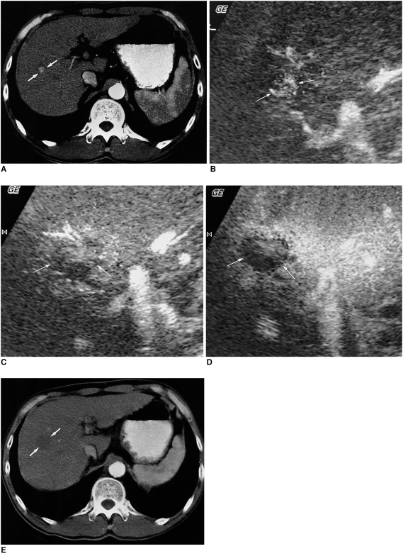
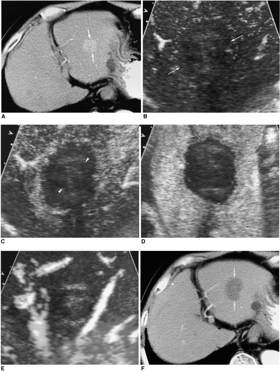
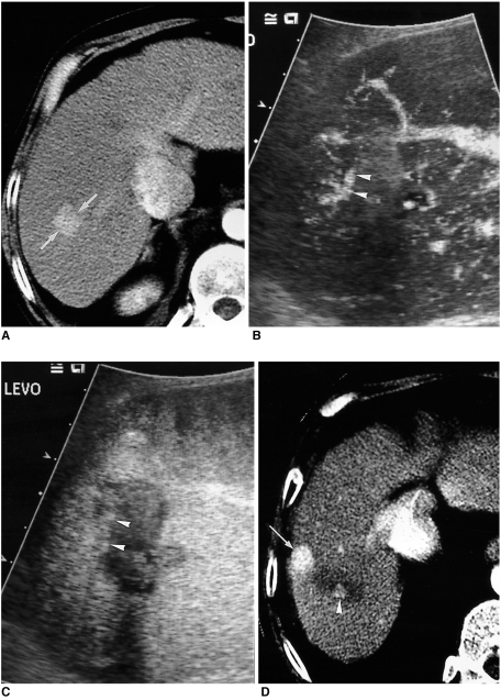
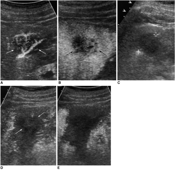
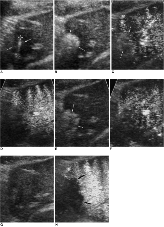
The early assessment of the therapeutic response after percutaneous radiofrequency (RF) ablation is important, in order to correctly decide whether further treatment is necessary. The residual unablated tumor is usually depicted on contrast-enhanced multiphase helical computed tomography (CT) as a focal enhancing structure during the arterial and portal venous phases. Contrast-enhanced color Doppler and power Doppler ultrasonography (US) have also been used to detect residual tumors. Contrast-enhanced gray-scale US, using a harmonic technology which has recently been introduced, allows for the detection of residual tumors after ablation, without any of the blooming or motion artifacts usually seen on contrast-enhanced color or power Doppler US. Based on our experience and reports in the literature, we consider that contrast-enhanced gray-scale harmonic US constitutes a reliable alternative to contrast-enhanced multiphase CT for the early evaluation of the therapeutic response to RF ablation for liver cancer. This technique was also useful in targeting any residual unablated tumors encountered during additional ablation.
经皮射频(RF)消融术后治疗反应的早期评估很重要,以便正确决定是否需要进一步治疗。残余未消融的肿瘤在多期螺旋CT增强扫描中,通常在动脉期和门静脉期表现为局灶性强化结构。彩色多普勒和能量多普勒超声造影(US)也已用于检测残余肿瘤。最近引入的使用谐波技术的灰阶超声造影,能够检测消融后的残余肿瘤,而不会出现彩色或能量多普勒超声造影中常见的伪像或运动伪影。基于我们的经验和文献报道,我们认为灰阶超声造影是多期CT增强扫描的可靠替代方法,可用于早期评估肝癌射频消融的治疗反应。该技术在引导额外消融过程中遇到的任何残余未消融肿瘤时也很有用。