Iwai Yoshiko, Terawaki Seigo, Ikegawa Masaya, Okazaki Taku, Honjo Tasuku
Department of Medical Chemistry, Graduate School of Medicine, Kyoto University, Yoshida, Sakyo-ku, Kyoto 606-8501, Japan.
J Exp Med. 2003 Jul 7;198(1):39-50. doi: 10.1084/jem.20022235.
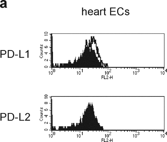
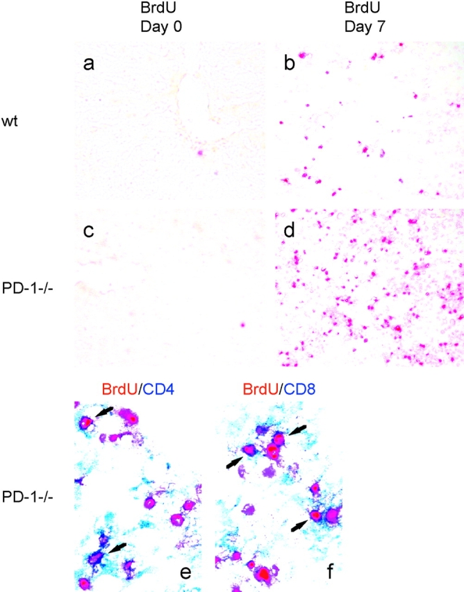
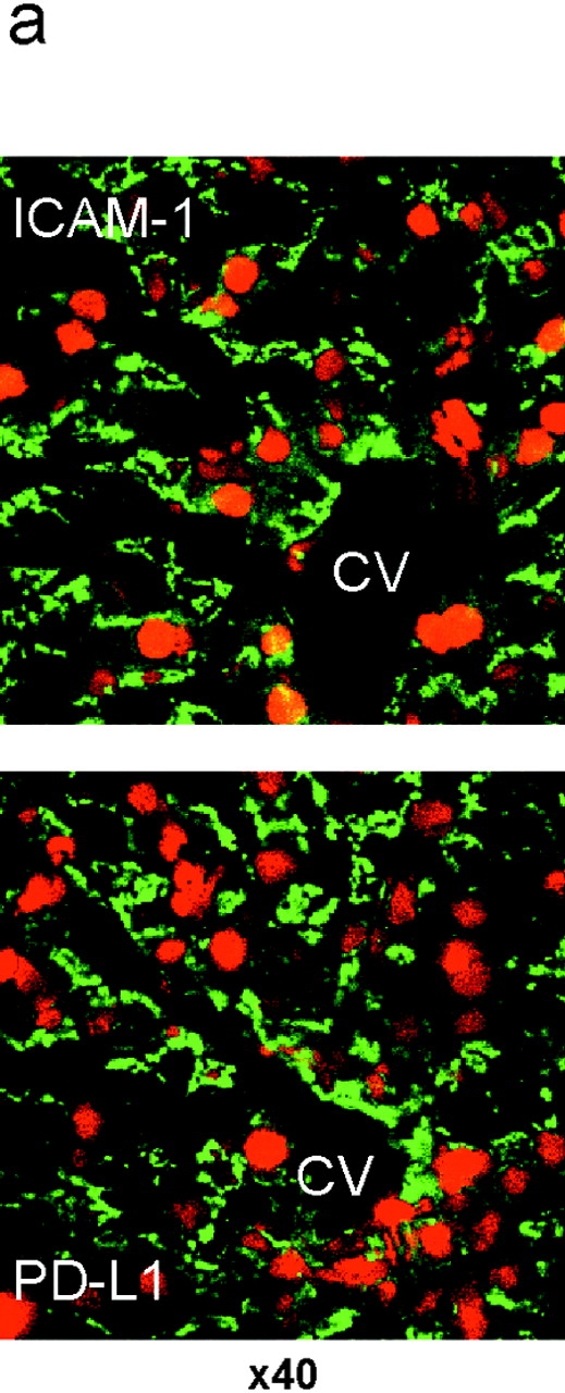
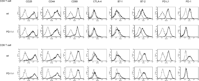
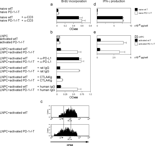
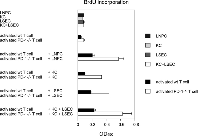
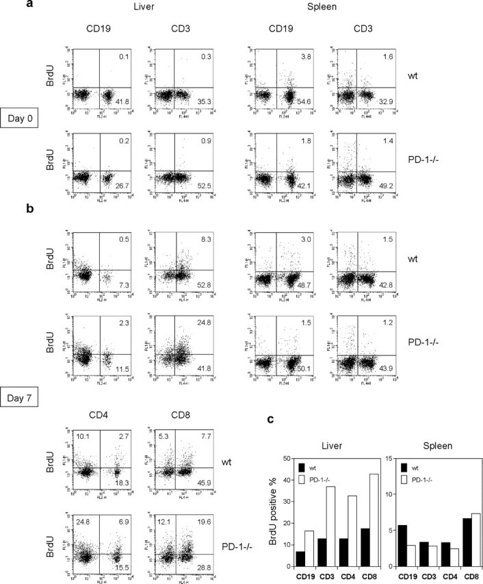
Unlike naive T cells, effector T cells can be activated by either T cell receptor signal or costimulatory signal alone and therefore the absence of costimulatory molecules on tissue cells cannot explain the tolerance mechanism at the effector phase. Here we report that PD-L1, the ligand for the immunoinhibitory receptor PD-1, was expressed on vascular endothelium in peripheral tissues. Liver nonparenchymal cells including sinusoidal endothelial cells and Kupffer cells constitutively expressed PD-L1 and inhibited proliferation and cell division of activated T cells expressing PD-1. The absence of PD-1 induced proliferation of effector T cells in the adenovirus-infected liver and resulted in rapid clearance of the virus. These results indicate that PD-1 plays an important role in T cell tolerance at the effector phase and the blockade of the PD-1 pathway can augment antiviral immunity.
与初始T细胞不同,效应T细胞可以仅被T细胞受体信号或共刺激信号激活,因此组织细胞上缺乏共刺激分子无法解释效应阶段的耐受机制。在此我们报告,免疫抑制受体PD-1的配体PD-L1在外周组织的血管内皮上表达。包括窦状内皮细胞和库普弗细胞在内的肝脏非实质细胞组成性表达PD-L1,并抑制表达PD-1的活化T细胞的增殖和细胞分裂。在腺病毒感染的肝脏中,缺乏PD-1会诱导效应T细胞增殖,并导致病毒的快速清除。这些结果表明,PD-1在效应阶段的T细胞耐受中起重要作用,阻断PD-1通路可以增强抗病毒免疫力。