Okafor Chukwuka C, Perreault-Micale Cynthia, Hajjar Roger J, Lebeche Djamel, Skiroman Klara, Jabbour George, Doye Angelia A, Lee Michael X, Laste Nancy, Gwathmey Judith K
Boston University Medical Center, Boston, USA.
BMC Physiol. 2003 Jul 21;3:6. doi: 10.1186/1472-6793-3-6.
Beta blocker treatment has emerged as an effective treatment modality for heart failure. Interestingly, beta-blockers can activate both pro-apoptotic and anti-apoptotic pathways. Nevertheless, the mechanism for improved cardiac function seen with beta-blocker treatment remains largely unknown. Carvedilol is a non-selective beta-blocker with alpha-receptor blockade and antioxidant properties. We therefore studied the impact of the effects of carvedilol in an animal model of end-stage heart failure.
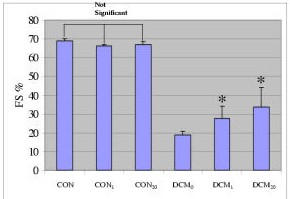
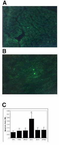
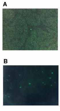
To test whether chronic treatment with beta-blockade decreases apoptosis, we treated myopathic turkeys with two dosages of carvedilol, 1 mg/kg (DCM1) and 20 mg/kg (DCM20), for four weeks and compared them to non-treated DCM animals (DCM0) and to control turkeys (CON). Echocardiographic measurements showed that non-treated DCM animals had a significantly lower fractional shortening (FS) when compared to CON (68.73 +/- 1.37 vs. 18.76 +/- 0.59%, p < 0.001). Both doses of carvedilol significantly improved FS (33.83 +/- 10.11 and 27.73 +/- 6.18% vs. 18.76 +/- 0.59% for untreated DCM, p < 0.001). DCM left ventricles were characterized by a higher percentage of apoptotic nuclei when compared to CON (5.64 +/- 0.49 vs. 1.72 +/- 0.12%, respectively p < 0.001). Both doses of carvedilol significantly reduced the number of apoptotic nuclei (2.32 +/- 0.23% and 2.36 +/-6% 1 mg and 20 mg/kg respectively).
Carvedilol improves ventricular function. Furthermore, treatment with carvedilol decreased the incidence of apoptosis in cardiac myocytes from failing hearts at both doses. These data suggest that the inhibition of apoptosis with carvedilol may lead to improvement in ventricular function and may underlie a beneficial effect of beta-blockade independent of heart rate lowering effects.
β受体阻滞剂治疗已成为心力衰竭的一种有效治疗方式。有趣的是,β受体阻滞剂可激活促凋亡和抗凋亡两条途径。然而,β受体阻滞剂治疗改善心脏功能的机制仍 largely 未知。卡维地洛是一种具有α受体阻滞作用和抗氧化特性的非选择性β受体阻滞剂。因此,我们在终末期心力衰竭动物模型中研究了卡维地洛作用的影响。
为测试β受体阻滞的长期治疗是否能减少细胞凋亡,我们用两种剂量的卡维地洛(1mg/kg,DCM1;20mg/kg,DCM20)对患心肌病的火鸡进行了四周治疗,并将它们与未治疗的扩张型心肌病(DCM)动物(DCM0)以及对照火鸡(CON)进行比较。超声心动图测量显示,与 CON 相比,未治疗的 DCM 动物的缩短分数(FS)显著更低(68.73±1.37 对 18.76±0.59%,p<0.001)。两种剂量的卡维地洛均显著改善了 FS(分别为 33.83±10.11 和 27.73±6.18%,而未治疗的 DCM 为 18.76±0.59%,p<0.001)。与 CON 相比,DCM 左心室的凋亡核百分比更高(分别为 5.64±0.49 对 1.72±0.12%,p<0.001)。两种剂量的卡维地洛均显著减少了凋亡核数量(分别为 2.32±0.23%和 2.36±6%,1mg/kg 和 20mg/kg)。
卡维地洛改善心室功能。此外,两种剂量的卡维地洛治疗均降低了衰竭心脏心肌细胞的凋亡发生率。这些数据表明,卡维地洛抑制细胞凋亡可能导致心室功能改善,且可能是β受体阻滞有益作用的基础,而与心率降低作用无关。