Alowami Salem, Troup Sandra, Al-Haddad Sahar, Kirkpatrick Iain, Watson Peter H
Department of Pathology and Molecular Medicine, McMaster University, Hamilton, Ontario, Canada.
Breast Cancer Res. 2003;5(5):R129-35. doi: 10.1186/bcr622. Epub 2003 Jul 23.
Mammographic density and certain histological changes in breast tissues are both risk factors for breast cancer. However, the relationship between these factors remains uncertain. Previous studies have focused on the histology of the epithelial changes, even though breast stroma is the major tissue compartment by volume. We have previously identified lumican and decorin as abundant small leucine-rich proteoglycans in breast stroma that show altered expression after breast tumorigenesis. In this study we have examined breast biopsies for a relationship between mammographic density and stromal alterations.
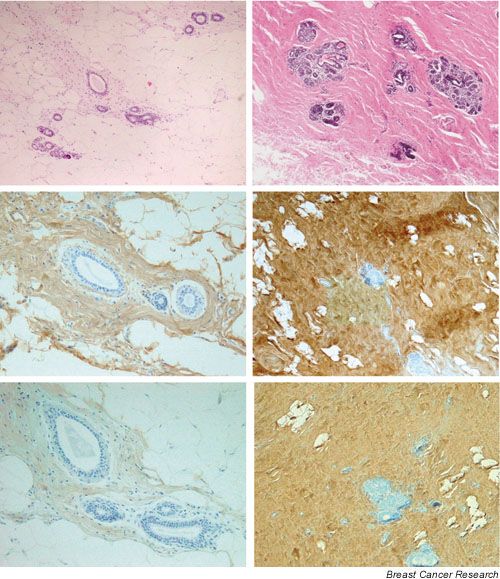
We reviewed mammograms from women aged 50-69 years who had enrolled in a provincial mammography screening program and had undergone an excision biopsy for an abnormality that was subsequently diagnosed as benign or pre-invasive breast disease. The overall mammographic density was classified into density categories. All biopsy tissue sections were reviewed and tissue blocks from excision margins distant from the diagnostic lesion were selected. Histological composition was assessed in sections stained with haematoxylin and eosin, and the expression of lumican and decorin was assessed by immunohistochemistry; both were quantified by semi-quantitative scoring.
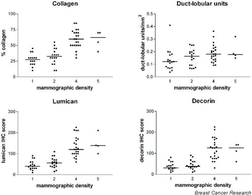
Tissue sections corresponding to regions of high in comparison with low mammographic density showed no significant difference in the density of ductal and lobular units but showed significantly higher collagen density and extent of fibrosis. Similarly, the expression of lumican and decorin was significantly increased.
Alteration in stromal composition is correlated with increased mammographic density. Although epithelial changes define the eventual pathway for breast cancer development, mammographic density might correspond more directly to alterations in stromal composition.
乳腺钼靶密度和乳腺组织的某些组织学变化均为乳腺癌的危险因素。然而,这些因素之间的关系仍不明确。既往研究主要关注上皮变化的组织学情况,尽管乳腺基质按体积计算是主要的组织成分。我们之前已确定,富含亮氨酸小分子蛋白聚糖(SLRP)的核心蛋白聚糖和饰胶蛋白聚糖在乳腺基质中含量丰富,且在乳腺肿瘤发生后表达发生改变。在本研究中,我们检查了乳腺活检样本,以探究乳腺钼靶密度与基质改变之间的关系。
我们回顾了年龄在50 - 69岁、参加省级乳腺钼靶筛查项目且因异常情况接受切除活检(随后被诊断为良性或乳腺原位癌)的女性的钼靶片。将整体乳腺钼靶密度分为不同密度类别。对所有活检组织切片进行复查,并选取距诊断性病变较远的切除边缘组织块。在苏木精 - 伊红染色切片中评估组织学组成,通过免疫组织化学评估核心蛋白聚糖和饰胶蛋白聚糖的表达;两者均通过半定量评分进行量化。
与乳腺钼靶低密度区域相对应的组织切片相比,高密度区域的导管和小叶单位密度无显著差异,但胶原密度和纤维化程度显著更高。同样,核心蛋白聚糖和饰胶蛋白聚糖的表达也显著增加。
基质组成的改变与乳腺钼靶密度增加相关。尽管上皮变化决定了乳腺癌发展的最终途径,但乳腺钼靶密度可能更直接地对应于基质组成的改变。