Subramanian S V, Satir B H
Department of Anatomy and Structural Biology, Albert Einstein College of Medicine, New York, NY 10461.
Proc Natl Acad Sci U S A. 1992 Dec 1;89(23):11297-301. doi: 10.1073/pnas.89.23.11297.
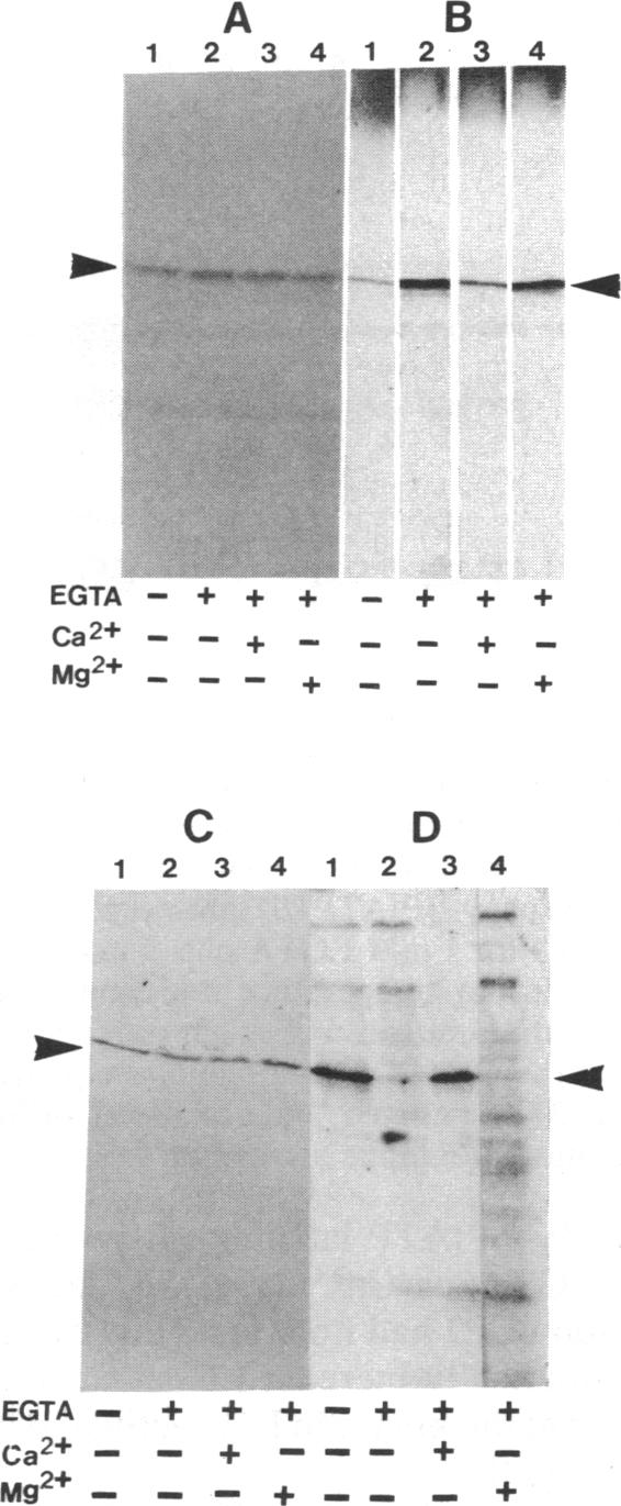
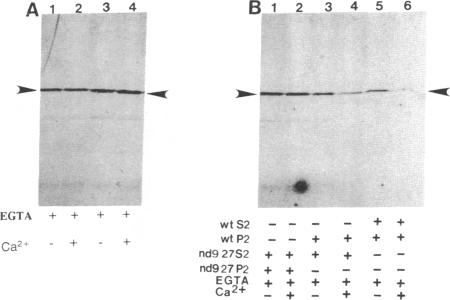
Parafusin, a cytosolic phosphoglycoprotein of M(r) 63,000, is dephosphorylated and rephosphorylated rapidly in a Ca(2+)-dependent manner upon stimulation of exocytosis in vivo in wild-type (wt) Paramecium. In contrast, the temperature-sensitive exocytosis mutant nd9, grown at the nonpermissive temperature (27 degrees C), does not exocytose or dephosphorylate parafusin upon stimulation in the presence of Ca2+; grown at the permissive temperature (18 degrees C), nd9 cells show a wt phenotype. Parafusin contains two types of phosphorylation sites: one where glucose 1-phosphate is added by an alpha-glucose-1-phosphate phosphotransferase and removed by a phosphodiesterase and one where phosphate from ATP is added directly to a serine residue by a protein kinase and removed by a phosphatase. We show here that, in cell fractions from wt Paramecium, both reactions can be carried out in vitro by using uridine(5'-[beta-[35S]thio])diphospho(1)-glucose (UDP[beta 35S]-Glc) and [gamma-32P]ATP, respectively. The characteristics of these pathways are different. Specifically, in the presence of Ca2+, the amount of UDP[beta 35S]-Glc label in parafusin is reduced. In contrast, identical labeling experiments with [gamma-32P]ATP show that Ca2+ enhances labeling of parafusin. Mg2+ had no appreciable effect on either labeling. Removal of the UDP[beta 35S]-Glc label on parafusin in the presence of Ca2+ correlates with the in vivo dephosphorylation seen upon exocytosis. Incubations with UDP[beta 35S]-Glc were then performed with homogenates and nd9 cell fractions grown at 27 degrees C under the ionic conditions used for wt cells. These labelings were not affected by Ca2+, in contrast to results from wt cells but in accord with those obtained earlier with nd9-27 mutant cells in vivo. Factors responsible for both dephosphorylation and Ca2+ sensitivity were found in the high-speed pellet (P2) in wt cells, suggesting that the putative phosphodiesterase is in this fraction and that the defect in the mutant nd9-27 residues in the Ca2+ activation of the phosphodiesterase. We conclude that the in vivo dephosphorylation of parafusin that occurs upon exocytosis is a dephosphoglucosylation due to removal of the alpha-glucose 1-phosphate and more generally that carbohydrates on cytoplasmic glycoproteins may be cyclically added and/or removed in response to extracellular stimuli.
副融合蛋白是一种分子量为63,000的胞质磷酸糖蛋白,在野生型草履虫体内,受到胞吐作用刺激后,它会以钙离子依赖的方式迅速发生去磷酸化和再磷酸化。相比之下,温度敏感型胞吐突变体nd9在非允许温度(27℃)下生长时,在钙离子存在的情况下受到刺激后不会发生胞吐作用,也不会使副融合蛋白去磷酸化;在允许温度(18℃)下生长时,nd9细胞表现出野生型表型。副融合蛋白含有两种磷酸化位点:一种是由α-葡萄糖-1-磷酸磷酸转移酶添加葡萄糖1-磷酸,并由磷酸二酯酶去除;另一种是由蛋白激酶将ATP中的磷酸直接添加到丝氨酸残基上,并由磷酸酶去除。我们在此表明,在野生型草履虫的细胞组分中,这两种反应分别可以通过使用尿苷(5'-[β-[35S]硫代])二磷酸(1)-葡萄糖(UDP[β35S]-Glc)和[γ-32P]ATP在体外进行。这些途径的特性不同。具体而言,在钙离子存在的情况下,副融合蛋白中UDP[β35S]-Glc标记的量减少。相反,用[γ-32P]ATP进行的相同标记实验表明,钙离子增强了副融合蛋白的标记。镁离子对这两种标记均无明显影响。在钙离子存在的情况下,副融合蛋白上UDP[β35S]-Glc标记的去除与胞吐作用时在体内观察到的去磷酸化相关。然后,在用于野生型细胞的离子条件下,用27℃下生长的匀浆和nd9细胞组分与UDP[β35S]-Glc进行孵育。与野生型细胞的结果相反,这些标记不受钙离子影响,但与之前在体内对nd9 - 27突变细胞获得的结果一致。在野生型细胞的高速沉淀(P2)中发现了负责去磷酸化和钙离子敏感性的因子,这表明假定的磷酸二酯酶在该组分中,并且突变体nd9 - 27的缺陷在于磷酸二酯酶的钙离子激活。我们得出结论,胞吐作用时发生的副融合蛋白的体内去磷酸化是由于α-葡萄糖1-磷酸的去除导致的去磷酸糖基化,更普遍地说,细胞质糖蛋白上的碳水化合物可能会响应细胞外刺激而循环添加和/或去除。