de Belder M A, Lovat L B, Tourikis L, Leech G, Camm A J
Department of Cardiological Sciences, St George's Hospital Medical School, London.
Br Heart J. 1992 Apr;67(4):297-303. doi: 10.1136/hrt.67.4.297.
To investigate the detection rate of cardiac sources of embolism by transoesophageal echocardiography in patients with focal cerebral ischaemic events and to relate the echocardiographic findings to other clinical findings.
Prospective study with blinded analysis of the echocardiographic data and subsequent comparison with the other clinical findings.
Regional cardiothoracic unit based in a teaching hospital.
131 consecutive patients with focal ischaemic cerebral events (49 with a transient ischaemic attack, 77 with a cerebrovascular accident, and five with a retinal arterial embolus) referred for echocardiography.
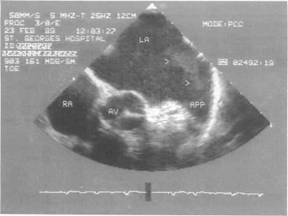
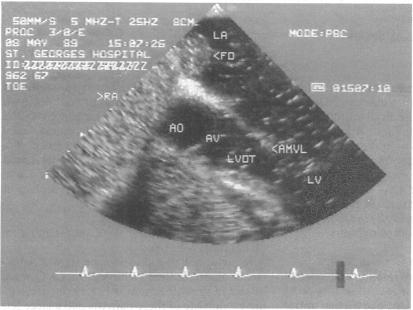
Full M mode, cross sectional, Doppler, and contrast echocardiography by both the precordial and transoesophageal techniques.
Precordial echocardiography detected a cardiac abnormality in 72 patients. Transoesophageal echocardiography confirmed all the precordial findings (except left ventricular hypertrophy, which at present cannot be defined with this technique) and detected other abnormalities in a further 20 patients (18 with potential right-to-left shunts and two with valve vegetations). It also showed spontaneous contrast echoes in 27 of 28 patients with a large left atrium and showed atrial thrombus in three. Cardiac abnormalities were clinically detected in 53 patients, all of which were confirmed or documented by echocardiography. In the 78 patients with no clinically detectable cardiac abnormality six had mitral valve prolapse and one had a regional wall motion defect (identified by precordial echocardiography) and 17 had potential right-to-left shunts (11 of which were identified only by transoesophageal echocardiography).
Transoesophageal echocardiography is more sensitive than precordial echocardiography in detecting potential sources of embolism in these patients. However, except for the detection of a potential right-to-left shunt, the yield in patients with no cardiac abnormality is low. Moreover, the abnormalities detected in those with previously detected cardiac disease merely confirm the clinical diagnosis. Patients with left atrial spontaneous contrast echoes may benefit from anticoagulation but this requires further study. Until more data are available on this feature and on the role of potential right-to-left shunts in this population, the contribution of echocardiography, precordial or transoesophageal, remains limited.
通过经食管超声心动图研究局灶性脑缺血事件患者心脏栓子来源的检出率,并将超声心动图结果与其他临床结果相关联。
前瞻性研究,对超声心动图数据进行盲法分析,随后与其他临床结果进行比较。
一家教学医院的区域心胸科。
131例连续的局灶性缺血性脑事件患者(49例短暂性脑缺血发作,77例脑血管意外,5例视网膜动脉栓塞)接受超声心动图检查。
采用心前区和经食管技术进行全M型、横截面、多普勒和对比超声心动图检查。
心前区超声心动图在72例患者中检测到心脏异常。经食管超声心动图证实了所有心前区检查结果(左心室肥厚除外,目前该技术无法定义),并在另外20例患者中检测到其他异常(18例有潜在的右向左分流,2例有瓣膜赘生物)。它还在28例左心房增大的患者中的27例中显示了自发对比回声,并在3例中显示了心房血栓。53例患者临床上检测到心脏异常,所有这些均通过超声心动图得到证实或记录。在78例临床上未检测到心脏异常的患者中,6例有二尖瓣脱垂,1例有节段性室壁运动缺损(通过心前区超声心动图确定),17例有潜在的右向左分流(其中11例仅通过经食管超声心动图确定)。
在检测这些患者潜在的栓子来源方面,经食管超声心动图比心前区超声心动图更敏感。然而,除了检测到潜在的右向左分流外,无心脏异常患者的检出率较低。此外,在先前检测到心脏病的患者中检测到的异常仅证实了临床诊断。左心房自发对比回声的患者可能从抗凝治疗中获益,但这需要进一步研究。在获得更多关于该特征以及潜在右向左分流在该人群中的作用的数据之前,心前区或经食管超声心动图的贡献仍然有限。