Black I W, Hopkins A P, Lee L C, Jacobson B M, Walsh W F
Department of Cardiovascular Medicine, Prince Henry/Prince of Wales Hospitals, Sydney, Australia.
Br Heart J. 1991 Oct;66(4):302-7. doi: 10.1136/hrt.66.4.302.
To determine the value of transoesophageal echocardiography in the assessment of selected patients at risk of cardiogenic embolism or after it.
Prospective comparison of the results of transoesophageal and transthoracic echocardiography. Transoesophageal echocardiography was performed with a 5 MHz single plane phased array transducer.
University teaching hospital.
100 patients referred for transoesophageal echocardiography after a cerebral ischaemic event or peripheral arterial embolism (n = 63), before percutaneous balloon dilatation of the mitral valve (n = 23), or before electrical cardioversion of atrial fibrillation (n = 14).
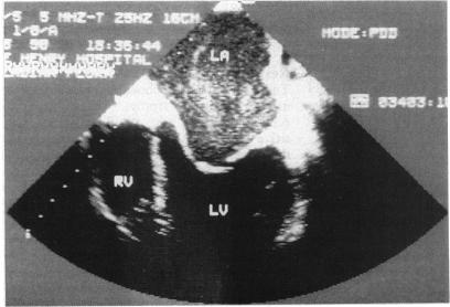
Transthoracic echocardiography showed potential sources of embolism in four patients including left ventricular thrombus in two patients (with one false positive), left atrial appendage thrombus (n = 1), and patent foramen ovale (n = 1). Transoesophageal echocardiography showed 59 potential embolic sources in 45 patients including left atrial spontaneous echo contrast (n = 33), left atrial appendage thrombus (n = 13), left ventricular thrombus (n = 5), patent foramen ovale (n = 3), left ventricular spontaneous echo contrast (n = 2), mitral valve prosthesis thrombus (n = 1), mitral valve prolapse (n = 1), and pronounced aortic atheroma (n = 1). Transoesophagal echocardiography showed potential embolic sources in 36/53 (68%) patients with atrial fibrillation compared with 9/47 (19%) patients in sinus rhythm. Percutaneous balloon dilatation of the mitral valve was performed without embolic complications in 18 patients without left atrial thrombi and in three patients with small fixed thrombi in the left atrial appendage. It was cancelled in two patients with large thrombi in the left atrial appendage. Cardioversion was performed without embolic complications in 14 patients without left atrial thrombi.
Transoesophageal echocardiography detects potential sources of embolism better than transthoracic echocardiography in selected patients at risk of cardiogenic embolism or after it.
确定经食管超声心动图在评估特定的心源性栓塞风险患者或栓塞发生后的患者中的价值。
经食管和经胸超声心动图结果的前瞻性比较。经食管超声心动图采用5MHz单平面相控阵探头进行检查。
大学教学医院。
100例患者,包括在发生脑缺血事件或外周动脉栓塞后接受经食管超声心动图检查的患者(n = 63)、在经皮二尖瓣球囊扩张术前的患者(n = 23)或在房颤电复律术前的患者(n = 14)。
经胸超声心动图显示4例患者存在潜在的栓塞源,包括2例左心室血栓患者(其中1例假阳性)、1例左心耳血栓患者和1例卵圆孔未闭患者。经食管超声心动图显示45例患者中有59个潜在的栓塞源,包括左心房自发显影(n = 33)、左心耳血栓(n = 13)、左心室血栓(n = 5)、卵圆孔未闭(n = 3)、左心室自发显影(n = 2)、二尖瓣人工瓣膜血栓(n = 1)、二尖瓣脱垂(n = 1)和明显的主动脉粥样硬化(n = 1)。经食管超声心动图显示,在36/53(68%)的房颤患者中存在潜在的栓塞源,而在窦性心律的患者中这一比例为9/47(19%)。18例无左心房血栓的患者和3例左心耳有小的固定血栓的患者在进行经皮二尖瓣球囊扩张术时未发生栓塞并发症。2例左心耳有大血栓的患者取消了手术。14例无左心房血栓的患者在进行电复律时未发生栓塞并发症。
在特定的心源性栓塞风险患者或栓塞发生后的患者中,经食管超声心动图比经胸超声心动图能更好地检测出潜在的栓塞源。