• 文献检索
  • 文档翻译
  • 深度研究
  • 学术资讯
  • Suppr Zotero 插件Zotero 插件
  • 邀请有礼
  • 套餐&价格
  • 历史记录
应用&插件
Suppr Zotero 插件Zotero 插件浏览器插件Mac 客户端Windows 客户端微信小程序
定价
高级版会员购买积分包购买API积分包
服务
文献检索文档翻译深度研究API 文档MCP 服务
关于我们
关于 Suppr公司介绍联系我们用户协议隐私条款
关注我们

Suppr 超能文献

核心技术专利:CN118964589B侵权必究
粤ICP备2023148730 号-1Suppr @ 2026

文献检索

告别复杂PubMed语法,用中文像聊天一样搜索,搜遍4000万医学文献。AI智能推荐,让科研检索更轻松。

立即免费搜索

文件翻译

保留排版,准确专业,支持PDF/Word/PPT等文件格式,支持 12+语言互译。

免费翻译文档

深度研究

AI帮你快速写综述,25分钟生成高质量综述,智能提取关键信息,辅助科研写作。

立即免费体验

新月柄杆菌的S层:通过电子显微镜进行三维图像重建和结构分析

The S-layer of Caulobacter crescentus: three-dimensional image reconstruction and structure analysis by electron microscopy.

作者信息

Smit J, Engelhardt H, Volker S, Smith S H, Baumeister W

机构信息

Max-Planck-Institut für Biochemie, Martinsreid bei Munich, Germany.

出版信息

J Bacteriol. 1992 Oct;174(20):6527-38. doi: 10.1128/jb.174.20.6527-6538.1992.

DOI:10.1128/jb.174.20.6527-6538.1992
PMID:1400205
原文链接:https://pmc.ncbi.nlm.nih.gov/articles/PMC207617/
Abstract

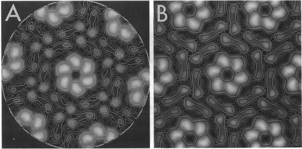
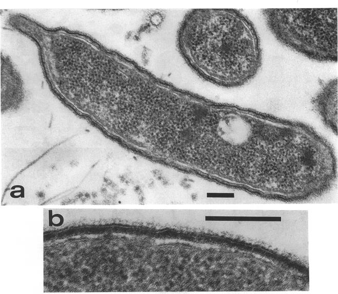
The regular surface protein structure (S-layer) of Caulobacter crescentus was analyzed by electron microscopy and three-dimensional image reconstruction to a resolution of 2 nm. Projections showed that the S-layer is an array of ring structures, each composed of six subunits that are arranged on a lattice with p6 symmetry. Three-dimensional reconstructions showed that the ring subunits were approximately rod-shaped structures and were perpendicular to the plane of the array, with a linker arm emanating from approximately the middle of the rod, accounting for the connections between the rings. The calculated subunit mass was ca. 100 kDa, very close to the size of RsaA (the protein known to be at least the predominant species in the S-layer) predicted from the DNA sequence of the rsaA gene. The core region of the rings creates an open pore 2.5 to 3.5 nm in diameter. The size of the gaps between the neighboring unit cells is in the same range, suggesting a uniform porosity predicted to exclude molecules larger than ca. 17 kDa. Attempts to remove membrane material from S-layer preparations with detergents revealed that the structure spontaneously rearranged into a mirror-image double layer. Negative-stain and thin-section electron microscopy examination of colonies of C. crescentus strains with a mutation in a surface molecule involved in the attachment of the S-layer showed that shed RsaA protein organized into large sheets. The sheets in turn organized into stacks that tended to accumulate near the upper surface of the colony. Image reconstruction indicated that these sheets were also precise mirror-image double layers, and thickness measurements obtained from thin sections were consistent with this finding. The sheets were absent when these mutant strains were grown without calcium, supporting other data that calcium is involved in attachment of the S-layer to a surface molecule and perhaps in subunit-subunit interactions. We propose that when the membrane is removed from S-layer fragments by detergents or the attachment-related surface molecule is absent, the attachment sites of the S-layer align precisely to form a double layer via a calcium interaction.

摘要

通过电子显微镜和三维图像重建技术对新月柄杆菌的规则表面蛋白结构(S层)进行了分析,分辨率达到2纳米。投影显示S层是由环状结构组成的阵列,每个环状结构由六个亚基组成,这些亚基以p6对称排列在晶格上。三维重建显示环状亚基近似为杆状结构,垂直于阵列平面,从杆的大约中间位置伸出一个连接臂,这解释了环之间的连接。计算得出的亚基质量约为100 kDa,与根据rsaA基因的DNA序列预测的RsaA(已知至少是S层中主要成分的蛋白质)大小非常接近。环的核心区域形成了一个直径为2.5至3.5纳米的开放孔。相邻晶胞之间的间隙大小在同一范围内,表明存在均匀的孔隙率,预计可排除大于约17 kDa的分子。用去污剂从S层制剂中去除膜材料的尝试表明,该结构会自发重排成镜像双层。对涉及S层附着的表面分子发生突变的新月柄杆菌菌株菌落进行的负染和超薄切片电子显微镜检查显示,脱落的RsaA蛋白组织成大片。这些片层进而组织成堆叠,倾向于在菌落的上表面附近积累。图像重建表明这些片层也是精确的镜像双层,从超薄切片获得的厚度测量结果与这一发现一致。当这些突变菌株在无钙条件下生长时,片层不存在,这支持了其他数据,即钙参与S层与表面分子的附着,也许还参与亚基 - 亚基相互作用。我们提出,当通过去污剂从S层片段中去除膜或不存在与附着相关的表面分子时,S层的附着位点会精确对齐,通过钙相互作用形成双层。

https://cdn.ncbi.nlm.nih.gov/pmc/blobs/2861/207617/347ade9d8567/jbacter00086-0229-a.jpg
https://cdn.ncbi.nlm.nih.gov/pmc/blobs/2861/207617/c2a5f068235f/jbacter00086-0220-a.jpg
https://cdn.ncbi.nlm.nih.gov/pmc/blobs/2861/207617/ead7659ffe48/jbacter00086-0221-a.jpg
https://cdn.ncbi.nlm.nih.gov/pmc/blobs/2861/207617/f58fa23495a2/jbacter00086-0222-a.jpg
https://cdn.ncbi.nlm.nih.gov/pmc/blobs/2861/207617/9536dacd99d0/jbacter00086-0223-a.jpg
https://cdn.ncbi.nlm.nih.gov/pmc/blobs/2861/207617/f4928f3a5be8/jbacter00086-0224-a.jpg
https://cdn.ncbi.nlm.nih.gov/pmc/blobs/2861/207617/c33ca01f7265/jbacter00086-0225-a.jpg
https://cdn.ncbi.nlm.nih.gov/pmc/blobs/2861/207617/99e1dc9077a8/jbacter00086-0226-a.jpg
https://cdn.ncbi.nlm.nih.gov/pmc/blobs/2861/207617/c8f1a54ee1df/jbacter00086-0227-a.jpg
https://cdn.ncbi.nlm.nih.gov/pmc/blobs/2861/207617/9694fa06867a/jbacter00086-0228-a.jpg
https://cdn.ncbi.nlm.nih.gov/pmc/blobs/2861/207617/347ade9d8567/jbacter00086-0229-a.jpg

相似文献

1
The S-layer of Caulobacter crescentus: three-dimensional image reconstruction and structure analysis by electron microscopy.新月柄杆菌的S层:通过电子显微镜进行三维图像重建和结构分析
J Bacteriol. 1992 Oct;174(20):6527-38. doi: 10.1128/jb.174.20.6527-6538.1992.
2
Characterization of mutants of Caulobacter crescentus defective in surface attachment of the paracrystalline surface layer.新月柄杆菌副晶质表层表面附着缺陷型突变体的特性分析
J Bacteriol. 1994 Oct;176(20):6312-23. doi: 10.1128/jb.176.20.6312-6323.1994.
3
Analysis of the intact surface layer of Caulobacter crescentus by cryo-electron tomography.利用冷冻电子断层成像术分析新月柄杆菌完整的表面层。
J Bacteriol. 2010 Nov;192(22):5855-65. doi: 10.1128/JB.00747-10. Epub 2010 Sep 10.
4
Linker mutagenesis of the Caulobacter crescentus S-layer protein: toward a definition of an N-terminal anchoring region and a C-terminal secretion signal and the potential for heterologous protein secretion.新月柄杆菌S层蛋白的接头诱变:对N端锚定区域和C端分泌信号的定义以及异源蛋白分泌的潜力
J Bacteriol. 1997 Feb;179(3):601-11. doi: 10.1128/jb.179.3.601-611.1997.
5
Cell-surface display of a Pseudomonas aeruginosa strain K pilin peptide within the paracrystalline S-layer of Caulobacter crescentus.在新月柄杆菌的准晶体S层内展示铜绿假单胞菌K型菌毛蛋白肽的细胞表面展示。
Mol Microbiol. 1997 Oct;26(2):277-88. doi: 10.1046/j.1365-2958.1997.5711932.x.
6
S-layer anchoring and localization of an S-layer-associated protease in Caulobacter crescentus.新月柄杆菌中S层锚定及一种S层相关蛋白酶的定位
J Bacteriol. 2007 Mar;189(6):2226-37. doi: 10.1128/JB.01690-06. Epub 2007 Jan 5.
7
Factors controlling in vitro recrystallization of the Caulobacter crescentus paracrystalline S-layer.控制新月柄杆菌副晶质表层体外重结晶的因素。
J Bacteriol. 1997 Oct;179(20):6349-54. doi: 10.1128/jb.179.20.6349-6354.1997.
8
Identification of lipopolysaccharide O antigen synthesis genes required for attachment of the S-layer of Caulobacter crescentus.新月柄杆菌S层附着所需的脂多糖O抗原合成基因的鉴定。
Microbiology (Reading). 2001 Jun;147(Pt 6):1451-1460. doi: 10.1099/00221287-147-6-1451.
9
Nucleotide sequence analysis of the gene encoding the Caulobacter crescentus paracrystalline surface layer protein.编码新月柄杆菌副晶质表层蛋白的基因的核苷酸序列分析。
Can J Microbiol. 1992 Mar;38(3):193-202. doi: 10.1139/m92-033.
10
The extreme N-terminus of the Caulobacter crescentus surface-layer protein directs export of passenger proteins from the cytoplasm but is not required for secretion of the native protein.新月柄杆菌表层蛋白的极端N端引导乘客蛋白从细胞质中输出,但对于天然蛋白的分泌并非必需。
Can J Microbiol. 1996 Jul;42(7):672-84. doi: 10.1139/m96-092.

引用本文的文献

1
Cell walls: a comparative view of the composition of cell surfaces of plants, algae, and microorganisms.细胞壁:植物、藻类和微生物细胞表面成分的比较观察
J Exp Bot. 2025 Jul 2;76(10):2614-2645. doi: 10.1093/jxb/erae512.
2
Composition and in situ structure of the cell envelope and surface layer.细胞包膜和表层的组成及原位结构。
Sci Adv. 2024 Dec 13;10(50):eadr8596. doi: 10.1126/sciadv.adr8596.
3
Cell cycle dependent coordination of surface layer biogenesis in Caulobacter crescentus.细胞周期依赖性协调新月柄杆菌表面层生物发生。

本文引用的文献

1
BIOLOGICAL PROPERTIES AND CLASSIFICATION OF THE CAULOBACTER GROUP.柄杆菌属的生物学特性与分类
Bacteriol Rev. 1964 Sep;28(3):231-95. doi: 10.1128/br.28.3.231-295.1964.
2
The correlation averaging of a regularly arranged bacterial cell envelope protein.规则排列的细菌细胞包膜蛋白的相关性平均
J Microsc. 1982 Aug;127(Pt 2):127-38. doi: 10.1111/j.1365-2818.1982.tb00405.x.
3
Three-dimensional reconstruction of imperfect two-dimensional crystals.不完美二维晶体的三维重建
Nat Commun. 2024 Apr 18;15(1):3355. doi: 10.1038/s41467-024-47529-5.
4
A Bayesian approach to single-particle electron cryo-tomography in RELION-4.0.基于 RELION-4.0 的单颗粒电子冷冻断层成像的贝叶斯方法。
Elife. 2022 Dec 5;11:e83724. doi: 10.7554/eLife.83724.
5
High-resolution mapping of metal ions reveals principles of surface layer assembly in Caulobacter crescentus cells.高分辨率金属离子图谱揭示了新月柄杆菌细胞表面层组装的原理。
Structure. 2022 Feb 3;30(2):215-228.e5. doi: 10.1016/j.str.2021.10.012. Epub 2021 Nov 19.
6
Engineering High-Yield Biopolymer Secretion Creates an Extracellular Protein Matrix for Living Materials.工程化高产生物聚合物分泌为生物材料构建细胞外蛋白质基质。
mSystems. 2021 Mar 23;6(2):e00903-20. doi: 10.1128/mSystems.00903-20.
7
Scratching the Surface: Bacterial Cell Envelopes at the Nanoscale. scratching the surface: bacterial cell envelopes at the nanoscale.
mBio. 2020 Feb 25;11(1):e03020-19. doi: 10.1128/mBio.03020-19.
8
In Situ Structure of an Intact Lipopolysaccharide-Bound Bacterial Surface Layer.完整脂多糖结合细菌表面层的原位结构。
Cell. 2020 Jan 23;180(2):348-358.e15. doi: 10.1016/j.cell.2019.12.006. Epub 2019 Dec 26.
9
Engineering the S-Layer of Caulobacter crescentus as a Foundation for Stable, High-Density, 2D Living Materials.改造新月柄杆菌的S层作为稳定、高密度二维生物材料的基础。
ACS Synth Biol. 2019 Jan 18;8(1):181-190. doi: 10.1021/acssynbio.8b00448. Epub 2019 Jan 7.
10
Locating macromolecules and determining structures inside bacterial cells using electron cryotomography.利用电子晶体学断层扫描技术定位细菌细胞内的大分子并确定其结构。
Biochim Biophys Acta Proteins Proteom. 2018 Sep;1866(9):973-981. doi: 10.1016/j.bbapap.2018.06.003. Epub 2018 Jun 13.
Ultramicroscopy. 1984;13(1-2):57-70. doi: 10.1016/0304-3991(84)90057-3.
4
Cloning of the major protein of the Caulobacter crescentus periodic surface layer: detection and characterization of the cloned peptide by protein expression assays.
J Bacteriol. 1984 Dec;160(3):1137-45. doi: 10.1128/jb.160.3.1137-1145.1984.
5
Periodic surface array in Caulobacter crescentus: fine structure and chemical analysis.新月柄杆菌中的周期性表面阵列:精细结构与化学分析
J Bacteriol. 1981 Jun;146(3):1135-50. doi: 10.1128/jb.146.3.1135-1150.1981.
6
Principles of organization in S layers.
J Mol Biol. 1986 Jan 20;187(2):251-3. doi: 10.1016/0022-2836(86)90232-9.
7
Three-dimensional structure of the regular surface layer (HPI layer) of Deinococcus radiodurans.耐辐射球菌规则表面层(HPI层)的三维结构。
J Mol Biol. 1986 Jan 20;187(2):241-50. doi: 10.1016/0022-2836(86)90231-7.
8
Nucleotide sequence analysis of the gene encoding the Deinococcus radiodurans surface protein, derived amino acid sequence, and complementary protein chemical studies.编码耐辐射球菌表面蛋白的基因的核苷酸序列分析、推导的氨基酸序列及互补蛋白化学研究。
J Bacteriol. 1987 Nov;169(11):5216-23. doi: 10.1128/jb.169.11.5216-5223.1987.
9
Three-dimensional structure of the regular tetragonal surface layer of Azotobacter vinelandii.
J Bacteriol. 1987 Nov;169(11):5008-15. doi: 10.1128/jb.169.11.5008-5015.1987.
10
Crystalline surface layers in procaryotes.原核生物中的晶体表面层。
J Bacteriol. 1988 Jul;170(7):2891-7. doi: 10.1128/jb.170.7.2891-2897.1988.