• 文献检索
  • 文档翻译
  • 深度研究
  • 学术资讯
  • Suppr Zotero 插件Zotero 插件
  • 邀请有礼
  • 套餐&价格
  • 历史记录
应用&插件
Suppr Zotero 插件Zotero 插件浏览器插件Mac 客户端Windows 客户端微信小程序
定价
高级版会员购买积分包购买API积分包
服务
文献检索文档翻译深度研究API 文档MCP 服务
关于我们
关于 Suppr公司介绍联系我们用户协议隐私条款
关注我们

Suppr 超能文献

核心技术专利:CN118964589B侵权必究
粤ICP备2023148730 号-1Suppr @ 2026

文献检索

告别复杂PubMed语法,用中文像聊天一样搜索,搜遍4000万医学文献。AI智能推荐,让科研检索更轻松。

立即免费搜索

文件翻译

保留排版,准确专业,支持PDF/Word/PPT等文件格式,支持 12+语言互译。

免费翻译文档

深度研究

AI帮你快速写综述,25分钟生成高质量综述,智能提取关键信息,辅助科研写作。

立即免费体验

秀丽隐杆线虫有丝分裂中心体中形态各异的微管末端。

Morphologically distinct microtubule ends in the mitotic centrosome of Caenorhabditis elegans.

作者信息

O'Toole Eileen T, McDonald Kent L, Mäntler Jana, McIntosh J Richard, Hyman Anthony A, Müller-Reichert Thomas

机构信息

Boulder Laboratory for 3-D Electron Microscopy of Cells, University of Colorado, 80309, USA.

出版信息

J Cell Biol. 2003 Nov 10;163(3):451-6. doi: 10.1083/jcb.200304035.

DOI:10.1083/jcb.200304035
PMID:14610052
原文链接:https://pmc.ncbi.nlm.nih.gov/articles/PMC2173630/
Abstract

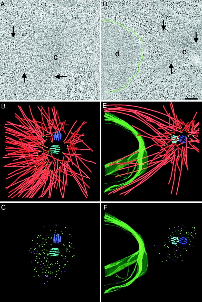
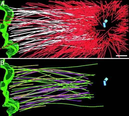
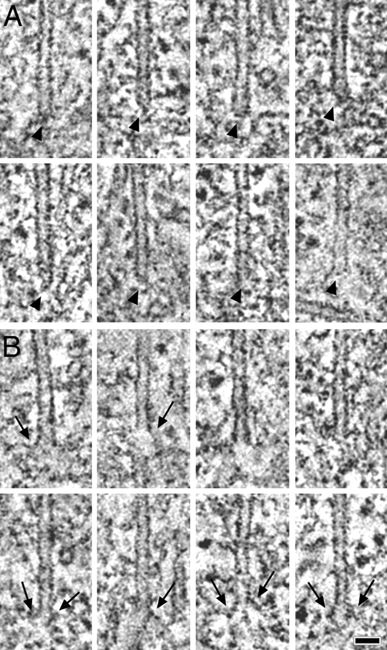
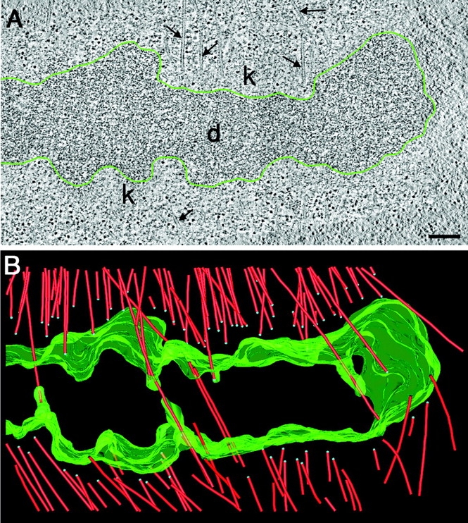
During mitosis, the connections of microtubules (MTs) to centrosomes and kinetochores are dynamic. From in vitro studies, it is known that the dynamic behavior of MTs is related to the structure of their ends, but we know little about the structure of MT ends in spindles. Here, we use high-voltage electron tomography to study the centrosome- and kinetochore-associated ends of spindle MTs in embryonic cells of the nematode, Caenorhabditis elegans. Centrosome-associated MT ends are either closed or open. Closed MT ends are more numerous and are uniformly distributed around the centrosome, but open ends are found preferentially on kinetochore-attached MTs. These results have structural implications for models of MT interactions with centrosomes.

摘要

在有丝分裂期间,微管(MTs)与中心体和动粒的连接是动态的。从体外研究可知,MTs的动态行为与其末端结构有关,但我们对纺锤体中MT末端的结构了解甚少。在这里,我们使用高压电子断层扫描技术来研究线虫秀丽隐杆线虫胚胎细胞中纺锤体MTs与中心体和动粒相关的末端。与中心体相关的MT末端要么是封闭的,要么是开放的。封闭的MT末端数量更多,且均匀分布在中心体周围,但开放末端优先出现在与动粒相连的MTs上。这些结果对MT与中心体相互作用的模型具有结构上的启示意义。

https://cdn.ncbi.nlm.nih.gov/pmc/blobs/7ebd/2173630/60466e33c394/200304035f5.jpg
https://cdn.ncbi.nlm.nih.gov/pmc/blobs/7ebd/2173630/bdaf4ec4e947/200304035f1.jpg
https://cdn.ncbi.nlm.nih.gov/pmc/blobs/7ebd/2173630/59d60e1cc5b6/200304035f2.jpg
https://cdn.ncbi.nlm.nih.gov/pmc/blobs/7ebd/2173630/56c970b75a34/200304035f3.jpg
https://cdn.ncbi.nlm.nih.gov/pmc/blobs/7ebd/2173630/5f5c56c92c9b/200304035f4.jpg
https://cdn.ncbi.nlm.nih.gov/pmc/blobs/7ebd/2173630/60466e33c394/200304035f5.jpg

相似文献

1
Morphologically distinct microtubule ends in the mitotic centrosome of Caenorhabditis elegans.秀丽隐杆线虫有丝分裂中心体中形态各异的微管末端。
J Cell Biol. 2003 Nov 10;163(3):451-6. doi: 10.1083/jcb.200304035.
2
Role of chromosomes in assembly of meiotic and mitotic spindles.染色体在减数分裂和有丝分裂纺锤体组装中的作用。
Prog Cell Cycle Res. 1997;3:271-84. doi: 10.1007/978-1-4615-5371-7_22.
3
Centromere Dysfunction Compromises Mitotic Spindle Pole Integrity.着丝粒功能障碍破坏有丝分裂纺锤体极的完整性。
Curr Biol. 2019 Sep 23;29(18):3072-3080.e5. doi: 10.1016/j.cub.2019.07.052. Epub 2019 Sep 5.
4
Cooperative mechanisms of mitotic spindle formation.有丝分裂纺锤体形成的协同机制。
J Cell Sci. 2007 May 15;120(Pt 10):1717-22. doi: 10.1242/jcs.03442.
5
C. elegans chromosomes connect to centrosomes by anchoring into the spindle network.秀丽隐杆线虫的染色体通过锚定在纺锤体网络中连接到中心体。
Nat Commun. 2017 May 11;8:15288. doi: 10.1038/ncomms15288.
6
The centrosome in vertebrates: more than a microtubule-organizing center.脊椎动物中的中心体:不仅仅是一个微管组织中心。
Trends Cell Biol. 2001 Oct;11(10):413-9. doi: 10.1016/s0962-8924(01)02085-2.
7
Genetic Control of Kinetochore-Driven Microtubule Growth in Mitosis.有丝分裂中动粒驱动的微管生长的遗传控制。
Cells. 2022 Jul 6;11(14):2127. doi: 10.3390/cells11142127.
8
The force-producing mechanism for centrosome separation during spindle formation in vertebrates is intrinsic to each aster.在脊椎动物纺锤体形成过程中,中心体分离的力产生机制是每个星体所固有的。
J Cell Biol. 1993 Jul;122(2):361-72. doi: 10.1083/jcb.122.2.361.
9
The elegans of spindle assembly.纺锤体组装的线虫。
Cell Mol Life Sci. 2010 Jul;67(13):2195-213. doi: 10.1007/s00018-010-0324-8. Epub 2010 Mar 26.
10
Polyglutamylated Tubulin Binding Protein C1orf96/CSAP Is Involved in Microtubule Stabilization in Mitotic Spindles.多聚谷氨酰胺化微管蛋白结合蛋白C1orf96/CSAP参与有丝分裂纺锤体中的微管稳定化过程。
PLoS One. 2015 Nov 12;10(11):e0142798. doi: 10.1371/journal.pone.0142798. eCollection 2015.

引用本文的文献

1
A germline-specific role for unconventional components of the γ-tubulin complex in Caenorhabditis elegans.生殖细胞特异性 γ-微管蛋白复合物非典型成分在秀丽隐杆线虫中的作用。
J Cell Sci. 2023 Jul 1;136(13). doi: 10.1242/jcs.260922. Epub 2023 Jul 11.
2
The Centrosome: Conclusions and Perspectives.中心体:结论与展望。
Cells. 2022 Dec 5;11(23):3931. doi: 10.3390/cells11233931.
3
MCRS1 modulates the heterogeneity of microtubule minus-end morphologies in mitotic spindles.MCRS1 调节有丝分裂纺锤体中微管负端形态的异质性。

本文引用的文献

1
Dynamics and mechanics of the microtubule plus end.微管正端的动力学与力学
Nature. 2003 Apr 17;422(6933):753-8. doi: 10.1038/nature01600.
2
SAS-4 is a C. elegans centriolar protein that controls centrosome size.SAS-4是一种控制中心体大小的秀丽隐杆线虫中心粒蛋白。
Cell. 2003 Feb 21;112(4):575-87. doi: 10.1016/s0092-8674(03)00117-x.
3
Chromosome-microtubule interactions during mitosis.有丝分裂期间的染色体-微管相互作用。
Mol Biol Cell. 2023 Jan 1;34(1):ar1. doi: 10.1091/mbc.E22-08-0306-T. Epub 2022 Nov 9.
4
Molecular architecture of the C. elegans centriole.秀丽隐杆线虫中心体的分子结构。
PLoS Biol. 2022 Sep 15;20(9):e3001784. doi: 10.1371/journal.pbio.3001784. eCollection 2022 Sep.
5
Three-dimensional structure of kinetochore-fibers in human mitotic spindles.人类有丝分裂纺锤体中着丝粒纤维的三维结构。
Elife. 2022 Jul 27;11:e75459. doi: 10.7554/eLife.75459.
6
Structural and Functional Insights into the Microtubule Organizing Centers of and spp.对XX菌和XX菌微管组织中心的结构与功能洞察
Microorganisms. 2021 Dec 3;9(12):2503. doi: 10.3390/microorganisms9122503.
7
A cryo-ET survey of microtubules and intracellular compartments in mammalian axons.哺乳动物轴突中微管和细胞内隔室的冷冻电镜调查。
J Cell Biol. 2022 Feb 7;221(2). doi: 10.1083/jcb.202103154. Epub 2021 Dec 8.
8
Microtubule-associated proteins promote microtubule generation in the absence of γ-tubulin in human colon cancer cells.微管相关蛋白在人结肠癌细胞中促进微管的生成,而无需 γ-微管蛋白。
J Cell Biol. 2021 Dec 6;220(12). doi: 10.1083/jcb.202104114. Epub 2021 Nov 15.
9
An extended DNA-free intranuclear compartment organizes centrosome microtubules in malaria parasites.一个扩展的无 DNA 核内隔室组织疟原虫中的中心体微管。
Life Sci Alliance. 2021 Sep 17;4(11). doi: 10.26508/lsa.202101199. Print 2021 Nov.
10
Centrosome instability: when good centrosomes go bad.中心体不稳定:好的中心体如何变得不稳定。
Cell Mol Life Sci. 2021 Nov;78(21-22):6775-6795. doi: 10.1007/s00018-021-03928-1. Epub 2021 Sep 2.
Annu Rev Cell Dev Biol. 2002;18:193-219. doi: 10.1146/annurev.cellbio.18.032002.132412. Epub 2002 Apr 2.
4
Cryomethods for thin section electron microscopy.用于超薄切片电子显微镜检查的冷冻方法。
Methods Enzymol. 2002;351:96-123. doi: 10.1016/s0076-6879(02)51843-7.
5
The kinetically dominant assembly pathway for centrosomal asters in Caenorhabditis elegans is gamma-tubulin dependent.秀丽隐杆线虫中心体星状体在动力学上占主导地位的组装途径是依赖γ-微管蛋白的。
J Cell Biol. 2002 May 13;157(4):591-602. doi: 10.1083/jcb.200202047.
6
Centrosome composition and microtubule anchoring mechanisms.中心体组成与微管锚定机制。
Curr Opin Cell Biol. 2002 Feb;14(1):25-34. doi: 10.1016/s0955-0674(01)00290-3.
7
Re-evaluating centrosome function.重新评估中心体功能。
Nat Rev Mol Cell Biol. 2001 Sep;2(9):688-98. doi: 10.1038/35089575.
8
HIM-10 is required for kinetochore structure and function on Caenorhabditis elegans holocentric chromosomes.HIM-10对于秀丽隐杆线虫全着丝粒染色体上动粒的结构和功能是必需的。
J Cell Biol. 2001 Jun 11;153(6):1227-38. doi: 10.1083/jcb.153.6.1227.
9
The C. elegans zyg-1 gene encodes a regulator of centrosome duplication with distinct maternal and paternal roles in the embryo.秀丽隐杆线虫的zyg-1基因编码一种中心体复制调节因子,在胚胎中具有不同的母源和父源作用。
Cell. 2001 May 18;105(4):547-58. doi: 10.1016/s0092-8674(01)00338-5.
10
Organellar relationships in the Golgi region of the pancreatic beta cell line, HIT-T15, visualized by high resolution electron tomography.通过高分辨率电子断层扫描观察胰腺β细胞系HIT-T15高尔基体区域的细胞器关系。
Proc Natl Acad Sci U S A. 2001 Feb 27;98(5):2399-406. doi: 10.1073/pnas.051631998.