Medical Theoretical Center (MTZ), TU Dresden, Fetscherstrasse 74, 01307, Dresden, Germany.
Cell Mol Life Sci. 2010 Jul;67(13):2195-213. doi: 10.1007/s00018-010-0324-8. Epub 2010 Mar 26.
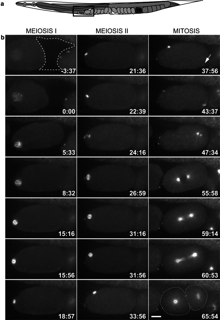
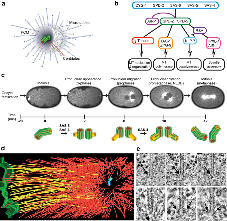
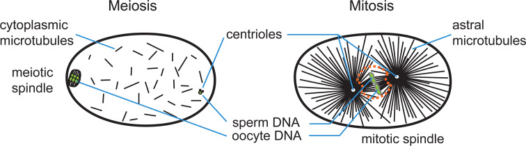
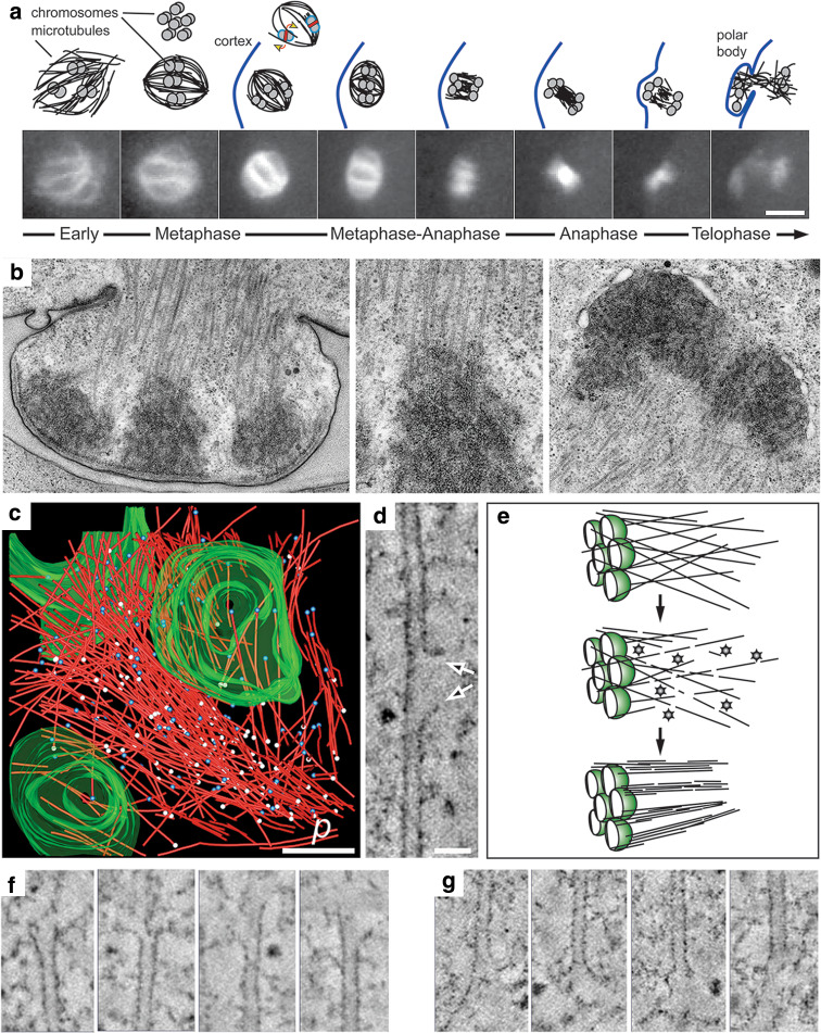
The Caenorhabditis elegans one-cell embryo is a powerful system in which to study microtubule organization because this large cell assembles both meiotic and mitotic spindles within the same cytoplasm over the course of 1 h in a stereotypical manner. The fertilized oocyte assembles two consecutive acentrosomal meiotic spindles that function to reduce the replicated maternal diploid set of chromosomes to a single-copy haploid set. The resulting maternal DNA then unites with the paternal DNA to form a zygotic diploid complement, around which a centrosome-based mitotic spindle forms. The early C. elegans embryo is amenable to live-cell imaging and electron tomography, permitting a detailed structural comparison of the meiotic and mitotic modes of spindle assembly.
秀丽隐杆线虫的单细胞胚胎是研究微管组织的强大系统,因为这个大细胞在 1 小时内以典型的方式在同一细胞质中组装减数分裂和有丝分裂纺锤体。受精卵组装两个连续的无中心体减数分裂纺锤体,其功能是将复制的母本二倍体染色体组减少到单个拷贝的单倍体组。然后,母本 DNA 与父本 DNA 结合形成合子二倍体补充物,围绕该补充物形成基于中心体的有丝分裂纺锤体。早期秀丽隐杆线虫胚胎适合活细胞成像和电子断层扫描,允许对减数分裂和有丝分裂纺锤体组装模式进行详细的结构比较。