Bauréus-Koch C, Nyberg G, Widegren B, Salford L G, Persson B R R
Department of Radiation Physics, Lund University Hospital, Klinikgatan 7, SE 221 85 Lund, Sweden.
Br J Cancer. 2004 Jan 12;90(1):48-54. doi: 10.1038/sj.bjc.6601467.
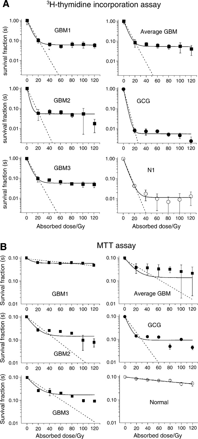
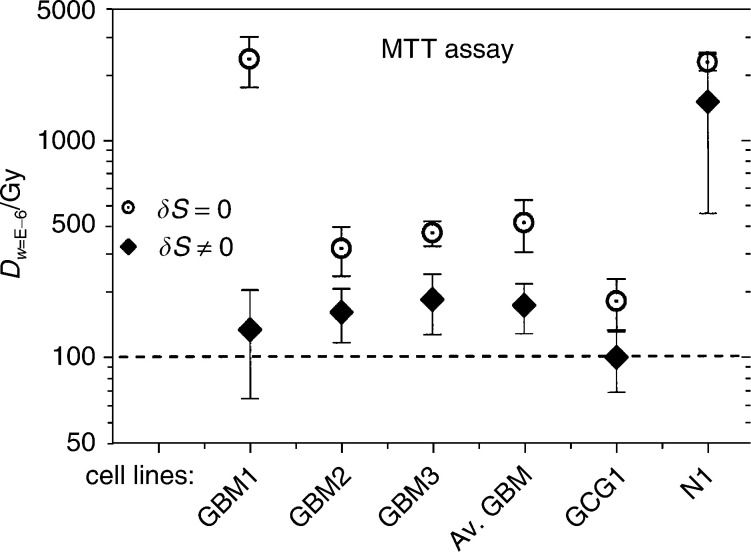
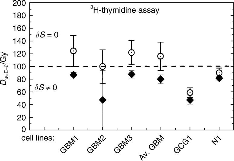
The aim is to investigate the radiosensitivity of noninfected cultured human glioma cells to ascertain that intracutaneously administered cells are viable enough to produce interferon-gamma but not able to proliferate. Cell cultures were established from five patients undergoing brain tumour surgery. By karyotyping, we found four malignant (three glioblastoma multiforme (GBM), one giant cell glioma) and one normal. The cells were irradiated with (137)Cs-gamma rays at absorbed dose levels of 0, 20, 40, 60, 80, 100 and 120 Gy. The fraction of viable cells was examined by MTT incorporation assay. The average of the data obtained from three GBM cell cultures was fitted to an exponential model. The parameters were: extrapolation number n=0.85+/-0.10, mean lethal dose D(0)=12.4+/-3.2 Gy and an additional uncertainty parameter deltaS=0.14+/-0.03. By setting deltaS=0, the corresponding values of the parameters were n=0.86+/-0.16 and D(0)=30.0+/-8.1 Gy. The rate of proliferation was examined by (3)H-thymidine incorporation. The average of the proliferation data obtained from three GBM cell cultures was fitted to an exponential model yielding n=0.943+/-0.005 and D(0)=5.8+/-0.5 Gy for deltaS=0.057+/-0.005, and by setting deltaS=0, n=1.00+/-0.02 and D(0)=8.4+/-1.6 Gy. No outgrowth of plated cells was observed after 4 weeks at an absorbed dose of 100 Gy. This absorbed dose is recommended for irradiation of 2 x 10(6) glioma cells used for clinical immunisation.
目的是研究未感染的培养人胶质瘤细胞的放射敏感性,以确定皮内注射的细胞有足够的活力产生γ干扰素,但不能增殖。从五名接受脑肿瘤手术的患者中建立细胞培养物。通过核型分析,我们发现四个恶性肿瘤(三个多形性胶质母细胞瘤(GBM),一个巨细胞胶质瘤)和一个正常细胞。细胞用137 Cs-γ射线以0、20、40、60、80、100和120 Gy的吸收剂量水平进行照射。通过MTT掺入试验检查存活细胞的比例。从三种GBM细胞培养物获得的数据平均值拟合到指数模型。参数为:外推数n = 0.85±0.10,平均致死剂量D(0)= 12.4±3.2 Gy和附加不确定参数δS = 0.14±0.03。通过设置δS = 0,参数的相应值为n = 0.86±0.16和D(0)= 30.0±8.1 Gy。通过3H-胸苷掺入检查增殖率。从三种GBM细胞培养物获得的增殖数据平均值拟合到指数模型,对于δS = 0.057±0.005,得到n = 0.943±0.005和D(0)= 5.8±0.5 Gy,并且通过设置δS = 0,n = 1.00±0.02和D(0)= 8.4±1.6 Gy。在100 Gy的吸收剂量下4周后未观察到接种细胞的生长。该吸收剂量推荐用于临床免疫所用的2×10^6个胶质瘤细胞的照射。