Goldsmith Cynthia S, Tatti Kathleen M, Ksiazek Thomas G, Rollin Pierre E, Comer James A, Lee William W, Rota Paul A, Bankamp Bettina, Bellini William J, Zaki Sherif R
Infectious Disease Pathology Activity, Division of Viral and Rickettsial Diseases, National Center for Infectious Diseases, Centers for Disease Control and Prevention, Atlanta, Georgia 30333, USA.
Emerg Infect Dis. 2004 Feb;10(2):320-6. doi: 10.3201/eid1002.030913.
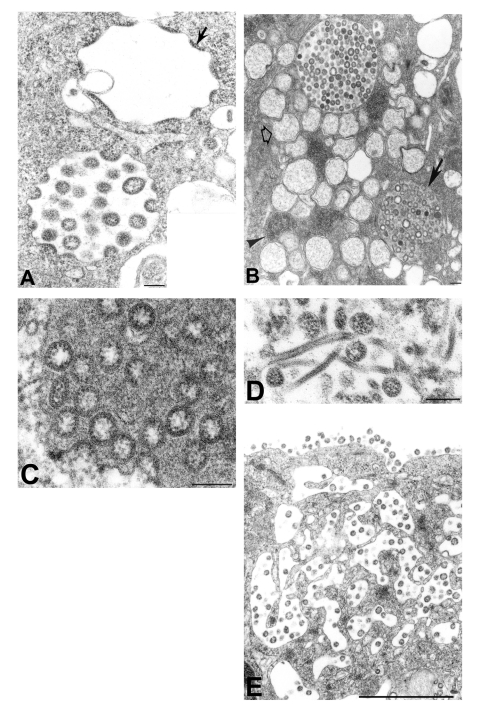
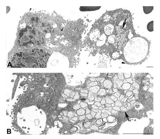
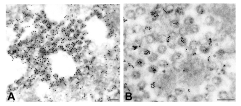
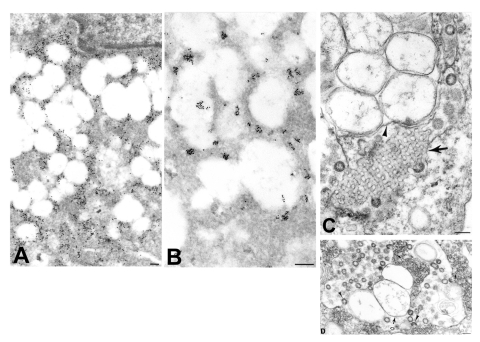
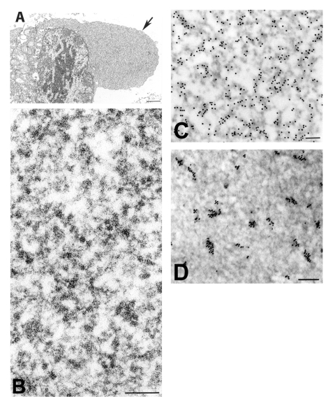
Severe acute respiratory syndrome (SARS) was first described during a 2002-2003 global outbreak of severe pneumonia associated with human deaths and person-to-person disease transmission. The etiologic agent was initially identified as a coronavirus by thin-section electron microscopic examination of a virus isolate. Virions were spherical, 78 nm in mean diameter, and composed of a helical nucleocapsid within an envelope with surface projections. We show that infection with the SARS-associated coronavirus resulted in distinct ultrastructural features: double-membrane vesicles, nucleocapsid inclusions, and large granular areas of cytoplasm. These three structures and the coronavirus particles were shown to be positive for viral proteins and RNA by using ultrastructural immunogold and in situ hybridization assays. In addition, ultrastructural examination of a bronchiolar lavage specimen from a SARS patient showed numerous coronavirus-infected cells with features similar to those in infected culture cells. Electron microscopic studies were critical in identifying the etiologic agent of the SARS outbreak and in guiding subsequent laboratory and epidemiologic investigations.
严重急性呼吸综合征(SARS)最初是在2002 - 2003年全球爆发的与人类死亡及人际间疾病传播相关的严重肺炎期间被描述的。通过对病毒分离株进行超薄切片电子显微镜检查,最初将病原体鉴定为一种冠状病毒。病毒粒子呈球形,平均直径78纳米,由包膜内的螺旋核衣壳和表面突起组成。我们发现,感染与SARS相关的冠状病毒会导致独特的超微结构特征:双膜囊泡、核衣壳包涵体以及细胞质的大颗粒区域。通过超微结构免疫金和原位杂交检测,这三种结构以及冠状病毒颗粒均显示病毒蛋白和RNA呈阳性。此外,对一名SARS患者的支气管肺泡灌洗标本进行超微结构检查,发现大量被冠状病毒感染的细胞,其特征与感染的培养细胞相似。电子显微镜研究对于确定SARS爆发的病原体以及指导后续的实验室和流行病学调查至关重要。