Tsujita-Kyutoku Miki, Yuri Takashi, Danbara Naoyuki, Senzaki Hideto, Kiyozuka Yasuhiko, Uehara Norihisa, Takada Hideho, Hada Takahiko, Miyazawa Teruo, Ogawa Yutaka, Tsubura Airo
Department of Pathology II, Kansai Medical University, Moriguchi, Osaka, Japan.
Breast Cancer Res. 2004;6(4):R291-9. doi: 10.1186/bcr789. Epub 2004 Apr 26.
The present study was conducted to examine the effect of conjugated docosahexaenoic acid (CDHA) on cell growth, cell cycle progression, mode of cell death, and expression of cell cycle regulatory and/or apoptosis-related proteins in KPL-1 human breast cancer cell line. This effect of CDHA was compared with that of docosahexaenoic acid (DHA).
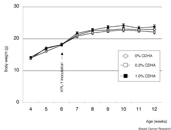
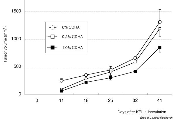
KPL-1 cell growth was assessed by colorimetric 3-(4,5-dimethylthiazol-2-yl)-2,5-diphenyltetrazolium bromide assay; cell cycle progression and mode of cell death were examined by flow cytometry; and levels of expression of p53, p21Cip1/Waf1, cyclin D1, Bax, and Bcl-2 proteins were examined by Western blotting analysis. In vivo tumor growth was examined by injecting KPL-1 cells subcutaneously into the area of the right thoracic mammary fat pad of female athymic mice fed a CDHA diet.
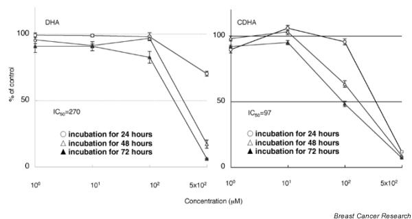
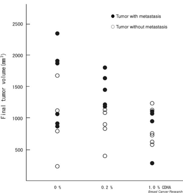
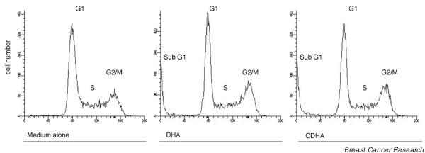
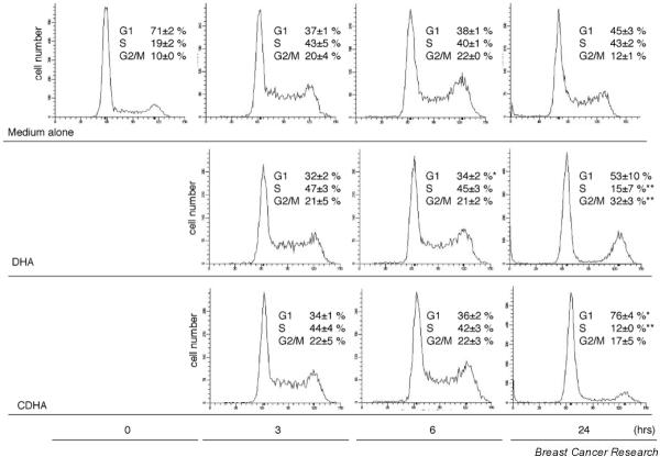
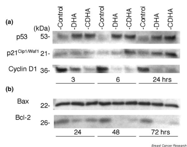
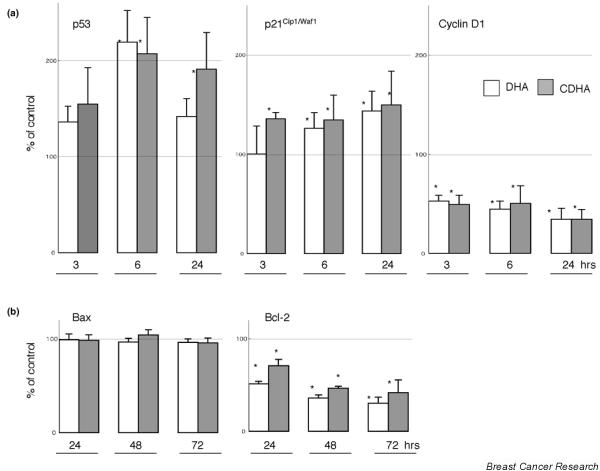
CDHA inhibited KPL-1 cells more effectively than did DHA (50% inhibitory concentration for 72 hours: 97 micromol/l and 270 micromol/l, respectively). With both CDHA and DHA growth inhibition was due to apoptosis, as indicated by the appearance of a sub-G1 fraction. The apoptosis cascade involved downregulation of Bcl-2 protein; Bax expression was unchanged. Cell cycle progression was due to G0/G1 arrest, which involved increased expression of p53 and p21Cip1/Waf1, and decreased expression of cyclin D1. CDHA modulated cell cycle regulatory proteins and apoptosis-related proteins in a manner similar to that of parent DHA. In the athymic mouse system 1.0% dietary CDHA, but not 0.2%, significantly suppressed growth of KPL-1 tumor cells; CDHA tended to decrease regional lymph node metastasis in a dose dependent manner.
CDHA inhibited growth of KPL-1 human breast cancer cells in vitro more effectively than did DHA. The mechanisms of action involved modulation of apoptosis cascade and cell cycle progression. Dietary CDHA at 1.0% suppressed KPL-1 cell growth in the athymic mouse system.
本研究旨在探讨共轭二十二碳六烯酸(CDHA)对KPL-1人乳腺癌细胞系的细胞生长、细胞周期进程、细胞死亡方式以及细胞周期调节蛋白和/或凋亡相关蛋白表达的影响。将CDHA的这种作用与二十二碳六烯酸(DHA)的作用进行比较。
通过比色法3-(4,5-二甲基噻唑-2-基)-2,5-二苯基四氮唑溴盐法评估KPL-1细胞的生长;通过流式细胞术检测细胞周期进程和细胞死亡方式;通过蛋白质印迹分析检测p53、p21Cip1/Waf1、细胞周期蛋白D1、Bax和Bcl-2蛋白的表达水平。通过将KPL-1细胞皮下注射到喂食CDHA饮食的雌性无胸腺小鼠右胸乳腺脂肪垫区域来检测体内肿瘤生长。
CDHA比DHA更有效地抑制KPL-1细胞(72小时的50%抑制浓度分别为97微摩尔/升和270微摩尔/升)。如亚G1期部分的出现所示,CDHA和DHA的生长抑制均归因于细胞凋亡。细胞凋亡级联反应涉及Bcl-2蛋白的下调;Bax表达未改变。细胞周期进程归因于G0/G1期阻滞,这涉及p53和p21Cip1/Waf1表达增加以及细胞周期蛋白D1表达降低。CDHA调节细胞周期调节蛋白和凋亡相关蛋白的方式与母体DHA相似。在无胸腺小鼠系统中,1.0%的饮食CDHA而非0.2%能显著抑制KPL-1肿瘤细胞的生长;CDHA倾向于以剂量依赖的方式减少区域淋巴结转移。
CDHA在体外比DHA更有效地抑制KPL-1人乳腺癌细胞的生长。作用机制涉及调节细胞凋亡级联反应和细胞周期进程。1.0%的饮食CDHA在无胸腺小鼠系统中抑制KPL-1细胞生长。