• 文献检索
  • 文档翻译
  • 深度研究
  • 学术资讯
  • Suppr Zotero 插件Zotero 插件
  • 邀请有礼
  • 套餐&价格
  • 历史记录
应用&插件
Suppr Zotero 插件Zotero 插件浏览器插件Mac 客户端Windows 客户端微信小程序
定价
高级版会员购买积分包购买API积分包
服务
文献检索文档翻译深度研究API 文档MCP 服务
关于我们
关于 Suppr公司介绍联系我们用户协议隐私条款
关注我们

Suppr 超能文献

核心技术专利:CN118964589B侵权必究
粤ICP备2023148730 号-1Suppr @ 2026

文献检索

告别复杂PubMed语法,用中文像聊天一样搜索,搜遍4000万医学文献。AI智能推荐,让科研检索更轻松。

立即免费搜索

文件翻译

保留排版,准确专业,支持PDF/Word/PPT等文件格式,支持 12+语言互译。

免费翻译文档

深度研究

AI帮你快速写综述,25分钟生成高质量综述,智能提取关键信息,辅助科研写作。

立即免费体验

产生睾酮的睾丸间质细胞的祖细胞被揭示。

Progenitor cells of the testosterone-producing Leydig cells revealed.

作者信息

Davidoff Michail S, Middendorff Ralf, Enikolopov Grigori, Riethmacher Dieter, Holstein Adolf F, Müller Dieter

机构信息

Institute of Anatomy, University of Hamburg, Germany.

出版信息

J Cell Biol. 2004 Dec 6;167(5):935-44. doi: 10.1083/jcb.200409107. Epub 2004 Nov 29.

DOI:10.1083/jcb.200409107
PMID:15569711
原文链接:https://pmc.ncbi.nlm.nih.gov/articles/PMC2172461/
Abstract

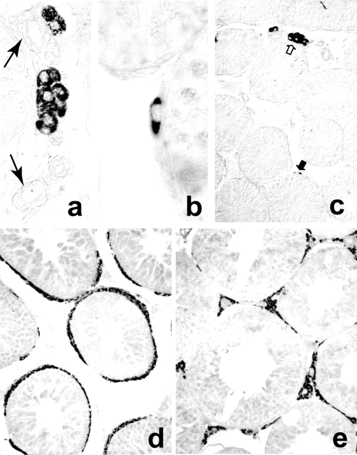
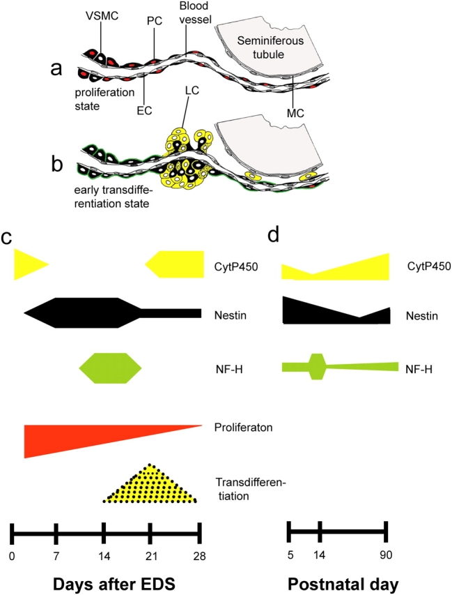
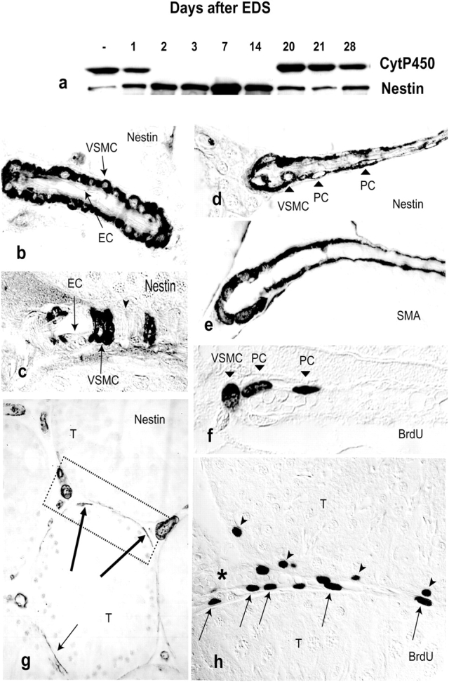
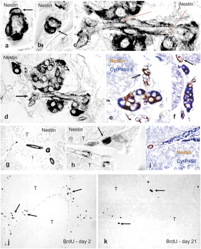
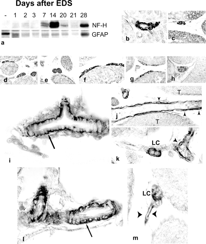
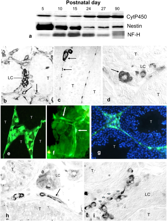
The cells responsible for production of the male sex hormone testosterone, the Leydig cells of the testis, are post-mitotic cells with neuroendocrine characteristics. Their origin during ontogeny and regeneration processes is still a matter of debate. Here, we show that cells of testicular blood vessels, namely vascular smooth muscle cells and pericytes, are the progenitors of Leydig cells. Resembling stem cells of the nervous system, the Leydig cell progenitors are characterized by the expression of nestin. Using an in vivo model to induce and monitor the synchronized generation of a completely new Leydig cell population in adult rats, we demonstrate specific proliferation of vascular progenitors and their subsequent transdifferentiation into steroidogenic Leydig cells which, in addition, rapidly acquire neuronal and glial properties. These findings, shown to be representative also for ontogenetic Leydig cell formation and for the human testis, provide further evidence that cellular components of blood vessels can act as progenitor cells for organogenesis and repair.

摘要

负责产生男性性激素睾酮的细胞,即睾丸间质细胞,是具有神经内分泌特征的有丝分裂后细胞。它们在个体发育和再生过程中的起源仍是一个有争议的问题。在这里,我们表明睾丸血管细胞,即血管平滑肌细胞和周细胞,是间质细胞的祖细胞。间质细胞祖细胞类似于神经系统的干细胞,其特征是巢蛋白的表达。利用体内模型诱导并监测成年大鼠全新间质细胞群体的同步生成,我们证明了血管祖细胞的特异性增殖及其随后向产生类固醇的间质细胞的转分化,此外,这些间质细胞还迅速获得神经元和神经胶质细胞特性。这些发现对于个体发育过程中间质细胞的形成以及人类睾丸也具有代表性,进一步证明血管的细胞成分可以作为器官发生和修复的祖细胞。

https://cdn.ncbi.nlm.nih.gov/pmc/blobs/d04c/2172461/3201faa6af36/200409107f6.jpg
https://cdn.ncbi.nlm.nih.gov/pmc/blobs/d04c/2172461/939e07b40c76/200409107f1.jpg
https://cdn.ncbi.nlm.nih.gov/pmc/blobs/d04c/2172461/eba5178a837e/200409107f2.jpg
https://cdn.ncbi.nlm.nih.gov/pmc/blobs/d04c/2172461/66620cf8a332/200409107f3.jpg
https://cdn.ncbi.nlm.nih.gov/pmc/blobs/d04c/2172461/7f421e55572c/200409107f4.jpg
https://cdn.ncbi.nlm.nih.gov/pmc/blobs/d04c/2172461/730851cce524/200409107f5.jpg
https://cdn.ncbi.nlm.nih.gov/pmc/blobs/d04c/2172461/3201faa6af36/200409107f6.jpg

相似文献

1
Progenitor cells of the testosterone-producing Leydig cells revealed.产生睾酮的睾丸间质细胞的祖细胞被揭示。
J Cell Biol. 2004 Dec 6;167(5):935-44. doi: 10.1083/jcb.200409107. Epub 2004 Nov 29.
2
The neuroendocrine Leydig cells and their stem cell progenitors, the pericytes.神经内分泌型睾丸间质细胞及其干细胞祖细胞,即周细胞。
Adv Anat Embryol Cell Biol. 2009;205:1-107.
3
Nestin, a neuroectodermal stem cell marker molecule, is expressed in Leydig cells of the human testis and in some specific cell types from human testicular tumours.巢蛋白是一种神经外胚层干细胞标记分子,在人类睾丸的间质细胞以及一些人类睾丸肿瘤的特定细胞类型中表达。
Cell Tissue Res. 2004 Jun;316(3):369-76. doi: 10.1007/s00441-003-0848-4. Epub 2004 May 4.
4
Hepatocyte growth factor is a mouse fetal Leydig cell terminal differentiation factor.肝细胞生长因子是一种诱导小鼠胎儿 Leydig 细胞终末分化的因子。
Biol Reprod. 2012 Dec 21;87(6):146. doi: 10.1095/biolreprod.112.104638. Print 2012 Jun.
5
Cell biology of Leydig cells in the testis.
Int Rev Cytol. 2004;233:181-241. doi: 10.1016/S0074-7696(04)33005-6.
6
Pituitary control of proliferation and differentiation of Leydig cells and their putative precursors in immature hypophysectomized rat testis.垂体对未成熟去垂体大鼠睾丸中Leydig细胞及其假定前体细胞增殖和分化的控制。
J Androl. 1996 Nov-Dec;17(6):639-50.
7
[Expression of P75NTR in the testis of nestin-GFP transgenic mice].[P75神经营养因子受体在巢蛋白绿色荧光蛋白转基因小鼠睾丸中的表达]
Zhonghua Nan Ke Xue. 2013 May;19(5):392-7.
8
Gene expression in rat leydig cells during development from the progenitor to adult stage: a cluster analysis.从祖细胞到成年阶段大鼠睾丸间质细胞发育过程中的基因表达:聚类分析
Biol Reprod. 2005 Jun;72(6):1405-15. doi: 10.1095/biolreprod.104.037499. Epub 2005 Feb 16.
9
De novo testosterone production in luteinizing hormone receptor knockout mice after transplantation of leydig stem cells.移植莱迪希干细胞后,促黄体生成素受体基因敲除小鼠中睾酮的从头合成。
Endocrinology. 2004 Sep;145(9):4011-5. doi: 10.1210/en.2003-1729. Epub 2004 May 3.
10
Glial fibrillary acidic protein-expressing neural progenitors give rise to immature neurons via early intermediate progenitors expressing both glial fibrillary acidic protein and neuronal markers in the adult hippocampus.胶质纤维酸性蛋白表达的神经祖细胞通过表达胶质纤维酸性蛋白和神经元标志物的早期中间祖细胞在成年海马体中产生未成熟神经元。
Neuroscience. 2010 Mar 10;166(1):241-51. doi: 10.1016/j.neuroscience.2009.12.026. Epub 2009 Dec 16.

引用本文的文献

1
Platelet-derived growth factor signaling in porcine testicular tubular somatic cells.猪睾丸曲细精管体细胞中的血小板源性生长因子信号传导
Cell Tissue Res. 2025 Aug 21. doi: 10.1007/s00441-025-04004-x.
2
NEDD4 Promotes Sertoli Cell Proliferation and Adult Leydig Cell Differentiation in the Murine Testis.NEDD4促进小鼠睾丸中支持细胞增殖和成年睾丸间质细胞分化。
Endocrinology. 2025 Jul 8;166(9). doi: 10.1210/endocr/bqaf115.
3
Efficient differentiation of human iPSCs into Leydig-like cells capable of long-term stable secretion of testosterone.

本文引用的文献

1
Nascent blood vessels in the skin arise from nestin-expressing hair-follicle cells.皮肤中的新生血管源自表达巢蛋白的毛囊细胞。
Proc Natl Acad Sci U S A. 2004 Sep 7;101(36):13291-5. doi: 10.1073/pnas.0405250101. Epub 2004 Aug 26.
2
One tissue, two fates: molecular genetic events that underlie testis versus ovary development.一种组织,两种命运:睾丸与卵巢发育背后的分子遗传事件。
Nat Rev Genet. 2004 Jul;5(7):509-21. doi: 10.1038/nrg1381.
3
Nestin, a neuroectodermal stem cell marker molecule, is expressed in Leydig cells of the human testis and in some specific cell types from human testicular tumours.
将人类诱导多能干细胞高效分化为能够长期稳定分泌睾酮的类睾丸间质细胞。
Stem Cell Reports. 2025 Feb 11;20(2):102392. doi: 10.1016/j.stemcr.2024.102392. Epub 2025 Jan 16.
4
A New Leydig Cell-Exclusive Cre Line Allows Lineage Tracing of Fetal and Adult Leydig Cell Populations in the Mouse.一种新的仅在睾丸间质细胞表达的Cre品系可用于对小鼠胎儿和成体睾丸间质细胞群体进行谱系追踪。
Endocrinology. 2025 Jan 6;166(2). doi: 10.1210/endocr/bqaf012.
5
Testicular sex cord-stromal tumors in mice with constitutive activation of PI3K and loss of Pten.PI3K组成性激活且Pten缺失的小鼠中的睾丸性索间质肿瘤
Carcinogenesis. 2025 Jan 20;46(1). doi: 10.1093/carcin/bgae077.
6
Age-related testosterone decline: mechanisms and intervention strategies.与年龄相关的睾酮下降:机制和干预策略。
Reprod Biol Endocrinol. 2024 Nov 14;22(1):144. doi: 10.1186/s12958-024-01316-5.
7
Effects of Leydig cell elimination on testicular interstitial cell populations: characterization by scRNA-seq and immunocytochemical techniques.Leydig 细胞消除对睾丸间质细胞群的影响:通过 scRNA-seq 和免疫细胞化学技术进行表征。
Front Endocrinol (Lausanne). 2024 Aug 20;15:1423801. doi: 10.3389/fendo.2024.1423801. eCollection 2024.
8
In vitro effects of uncarboxylated osteocalcin on buffalo Leydig cell steroidogenesis.体外非羧化骨钙素对水牛睾丸间质细胞类固醇生成的影响。
Vet Res Commun. 2024 Jun;48(3):1423-1433. doi: 10.1007/s11259-024-10320-4. Epub 2024 Feb 2.
9
The role of HIF-1α-mediated autophagy in ionizing radiation-induced testicular injury.低氧诱导因子 1α 介导体细胞自噬在电离辐射诱导睾丸损伤中的作用。
J Mol Histol. 2023 Oct;54(5):439-451. doi: 10.1007/s10735-023-10153-6. Epub 2023 Sep 20.
10
Distribution, contribution and regulation of nestin cells.巢蛋白细胞的分布、贡献和调控。
J Adv Res. 2024 Jul;61:47-63. doi: 10.1016/j.jare.2023.08.013. Epub 2023 Aug 28.
巢蛋白是一种神经外胚层干细胞标记分子,在人类睾丸的间质细胞以及一些人类睾丸肿瘤的特定细胞类型中表达。
Cell Tissue Res. 2004 Jun;316(3):369-76. doi: 10.1007/s00441-003-0848-4. Epub 2004 May 4.
4
Abundant progenitor cells in the adventitia contribute to atherosclerosis of vein grafts in ApoE-deficient mice.外膜中丰富的祖细胞促成了载脂蛋白E缺陷小鼠静脉移植物的动脉粥样硬化。
J Clin Invest. 2004 May;113(9):1258-65. doi: 10.1172/JCI19628.
5
Lost in transdifferentiation.迷失于转分化过程中。
J Clin Invest. 2004 May;113(9):1249-51. doi: 10.1172/JCI21761.
6
Early contribution of pericytes to angiogenic sprouting and tube formation.周细胞对血管生成芽生和管形成的早期贡献。
Angiogenesis. 2003;6(3):241-9. doi: 10.1023/B:AGEN.0000021401.58039.a9.
7
Neural stem and progenitor cells in nestin-GFP transgenic mice.巢蛋白绿色荧光蛋白转基因小鼠中的神经干细胞和祖细胞。
J Comp Neurol. 2004 Feb 9;469(3):311-24. doi: 10.1002/cne.10964.
8
The glial identity of neural stem cells.神经干细胞的神经胶质细胞特性
Nat Neurosci. 2003 Nov;6(11):1127-34. doi: 10.1038/nn1144. Epub 2003 Oct 28.
9
Roles of PDGF in animal development.血小板衍生生长因子在动物发育中的作用。
Development. 2003 Oct;130(20):4769-84. doi: 10.1242/dev.00721.
10
Blood vessels and nerves: common signals, pathways and diseases.血管与神经:共同的信号、通路及疾病
Nat Rev Genet. 2003 Sep;4(9):710-20. doi: 10.1038/nrg1158.