Malaspina Dolores, Harkavy-Friedman Jill, Corcoran Cheryl, Mujica-Parodi Lilianne, Printz David, Gorman Jack M, Van Heertum Ronald
New York State Psychiatric Institute, Columbia University, 1051 Riverside Drive, New York, NY 10032, USA.
Biol Psychiatry. 2004 Dec 15;56(12):931-7. doi: 10.1016/j.biopsych.2004.09.013.
Schizophrenia is etiologically heterogeneous. It is anticipated, but unproven, that subgroups will differ in neuropathology and that neuroimaging may reveal these differences. The optimal imaging condition may be at rest, where greater variability is observed than during cognitive tasks, which more consistently reveal hypofrontality. We previously demonstrated symptom and physiologic differences between familial and sporadic schizophrenia patients and hypothesized that the groups would show different resting regional cerebral blood flow (rCBF) patterns.
Ten familial and sixteen sporadic schizophrenia patients and nine comparison subjects had single photon emission computed tomography imaging during passive visual fixation. Images were spatially normalized into Talairach coordinates and analyzed for group rCBF differences using SPM with a Z value threshold of 2.80, p < .001.
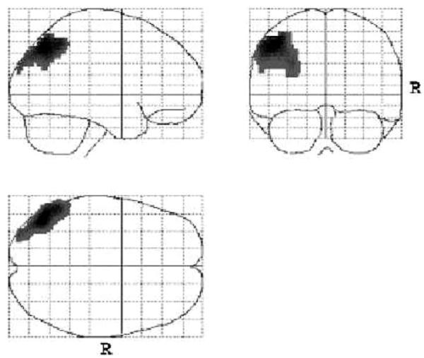
The subgroups had similar age, gender, illness duration, and medication treatment. Sporadic patients had hypofrontality (anterior cingulate, paracingulate cortices, left dorsolateral and inferior-orbitofrontal), whereas familial patients had left temporoparietal hypoperfusion; all of these regions show resting activity in healthy subjects. Both groups hyperperfused the cerebellum/pons and parahippocampal gyrus; additional hyperperfusion for sporadic patients was observed in the fusiform; familial patients also hyperperfused the hippocampus, dentate, uncus, amygdala, thalamus, and putamen.
Familial and sporadic schizophrenia patients had different resting rCBF profiles, supporting the hypothesis that certain subgroups have distinct neural underpinnings. Different neuropathologic processes among subgroups of schizophrenia patients may account for the prior contradictory results of resting imaging studies.
精神分裂症在病因上具有异质性。虽然预计亚组在神经病理学上会有所不同,但尚未得到证实,并且神经影像学可能会揭示这些差异。最佳成像条件可能是在静息状态下,此时观察到的变异性比认知任务期间更大,认知任务更一致地揭示前额叶功能低下。我们之前证明了家族性和散发性精神分裂症患者在症状和生理方面存在差异,并假设这两组患者会表现出不同的静息区域脑血流(rCBF)模式。
10名家族性精神分裂症患者、16名散发性精神分裂症患者和9名对照受试者在被动视觉注视期间进行了单光子发射计算机断层扫描成像。图像在空间上被标准化为Talairach坐标,并使用统计参数映射(SPM)分析组间rCBF差异,Z值阈值为2.80,p <.001。
亚组在年龄、性别、病程和药物治疗方面相似。散发性患者存在前额叶功能低下(前扣带回、旁扣带回皮质、左侧背外侧和眶额下回),而家族性患者左侧颞顶叶灌注不足;所有这些区域在健康受试者中均表现出静息活动。两组患者小脑/脑桥和海马旁回均存在血流灌注增加;散发性患者在梭状回还观察到额外的血流灌注增加;家族性患者在海马、齿状回、钩回、杏仁核、丘脑和壳核也存在血流灌注增加。
家族性和散发性精神分裂症患者具有不同的静息rCBF模式,支持了某些亚组具有不同神经基础的假设。精神分裂症患者亚组之间不同的神经病理过程可能解释了先前静息成像研究结果的矛盾之处。