Annaix V, Bouchara J P, Larcher G, Chabasse D, Tronchin G
Laboratoire de Parasitologie-Mycologie, Centre Hospitalier Régional et Universitaire, Angers, France.
Infect Immun. 1992 May;60(5):1747-55. doi: 10.1128/iai.60.5.1747-1755.1992.
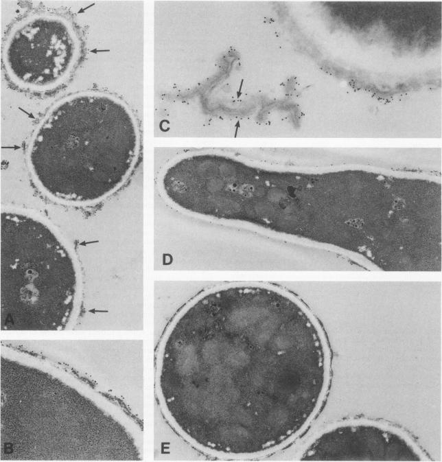
The interaction of purified human fibrinogen with Aspergillus fumigatus conidia was investigated by immunofluorescence and electron microscopy and binding assays with radiolabeled proteins. We described the localization of the binding sites on the A. fumigatus conidia and on the fibrinogen molecule and determined the binding characteristics. Immunofluorescence revealed that the fixation of purified fibrinogen was selectively associated with conidia and suggested a role for the D domains of the fibrinogen molecule. Binding assays performed with 125I-radiolabeled proteins confirmed that binding sites were located specifically in the D domains. No reaction could be detected with fragment E. The binding of 125I-fragment D to conidia was time dependent, saturable, and specific. Scatchard analysis of the data revealed an average of 1,200 binding sites per conidium, and an apparent dissociation constant (Kd) of 2.2 x 10(-9) M was estimated. Pretreatment of the cells with proteolytic enzymes or heat abolished binding, demonstrating the protein nature of the binding sites. Ultrastructural localization of the fungal receptors was determined by transmission electron microscopy. Labeling appeared to be associated with the outer electron-dense layer of the conidial wall and progressively decreased during the germination process. Labeling of thin sections with fragment D and an antifibrinogen immune serum revealed that binding sites also lay in the inner part of the wall and in vacuoles. These results indicate the presence at the conidial surface of specific receptors for fibrinogen which could act as mediators of conidial adherence to host tissues.
通过免疫荧光、电子显微镜以及放射性标记蛋白结合试验,研究了纯化的人纤维蛋白原与烟曲霉分生孢子之间的相互作用。我们描述了烟曲霉分生孢子和纤维蛋白原分子上结合位点的定位,并确定了结合特性。免疫荧光显示,纯化纤维蛋白原的固定与分生孢子选择性相关,提示纤维蛋白原分子的D结构域起作用。用¹²⁵I放射性标记蛋白进行的结合试验证实,结合位点特异性位于D结构域。用片段E未检测到反应。¹²⁵I-片段D与分生孢子的结合具有时间依赖性、饱和性和特异性。对数据进行Scatchard分析显示,每个分生孢子平均有1200个结合位点,估计表观解离常数(Kd)为2.2×10⁻⁹M。用蛋白水解酶或加热预处理细胞可消除结合,表明结合位点的蛋白质性质。通过透射电子显微镜确定真菌受体的超微结构定位。标记似乎与分生孢子壁的外部电子致密层相关,并且在萌发过程中逐渐减少。用片段D和抗纤维蛋白原免疫血清对薄片进行标记显示,结合位点也位于壁的内部和液泡中。这些结果表明,在分生孢子表面存在纤维蛋白原的特异性受体,其可作为分生孢子黏附于宿主组织的介质。