• 文献检索
  • 文档翻译
  • 深度研究
  • 学术资讯
  • Suppr Zotero 插件Zotero 插件
  • 邀请有礼
  • 套餐&价格
  • 历史记录
应用&插件
Suppr Zotero 插件Zotero 插件浏览器插件Mac 客户端Windows 客户端微信小程序
定价
高级版会员购买积分包购买API积分包
服务
文献检索文档翻译深度研究API 文档MCP 服务
关于我们
关于 Suppr公司介绍联系我们用户协议隐私条款
关注我们

Suppr 超能文献

核心技术专利:CN118964589B侵权必究
粤ICP备2023148730 号-1Suppr @ 2026

文献检索

告别复杂PubMed语法,用中文像聊天一样搜索,搜遍4000万医学文献。AI智能推荐,让科研检索更轻松。

立即免费搜索

文件翻译

保留排版,准确专业,支持PDF/Word/PPT等文件格式,支持 12+语言互译。

免费翻译文档

深度研究

AI帮你快速写综述,25分钟生成高质量综述,智能提取关键信息,辅助科研写作。

立即免费体验

利用几何背景均衡和阈值选择自动检测黄斑玻璃膜疣。

Automated detection of macular drusen using geometric background leveling and threshold selection.

作者信息

Smith R Theodore, Chan Jackie K, Nagasaki Takayuki, Ahmad Umer F, Barbazetto Irene, Sparrow Janet, Figueroa Marta, Merriam Joanna

机构信息

Edward S. Harkness Eye Institute, New York Presbyterian Medical Center, New York, NY 10032, USA.

出版信息

Arch Ophthalmol. 2005 Feb;123(2):200-6. doi: 10.1001/archopht.123.2.200.

DOI:10.1001/archopht.123.2.200
PMID:15710816
原文链接:https://pmc.ncbi.nlm.nih.gov/articles/PMC2884376/
Abstract

BACKGROUND

Age-related macular degeneration (ARMD) is the most prevalent cause of visual loss in patients older than 60 years in the United States. Observation of drusen is the hallmark finding in the clinical evaluation of ARMD.

OBJECTIVES

To segment and quantify drusen found in patients with ARMD using image analysis and to compare the efficacy of image analysis segmentation with that of stereoscopic manual grading of drusen.

DESIGN

Retrospective study.

SETTING

University referral center.Patients Photographs were randomly selected from an available database of patients with known ARMD in the ongoing Columbia University Macular Genetics Study. All patients were white and older than 60 years.

INTERVENTIONS

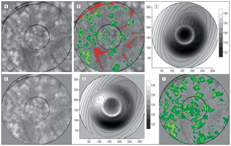
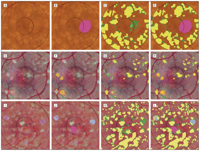
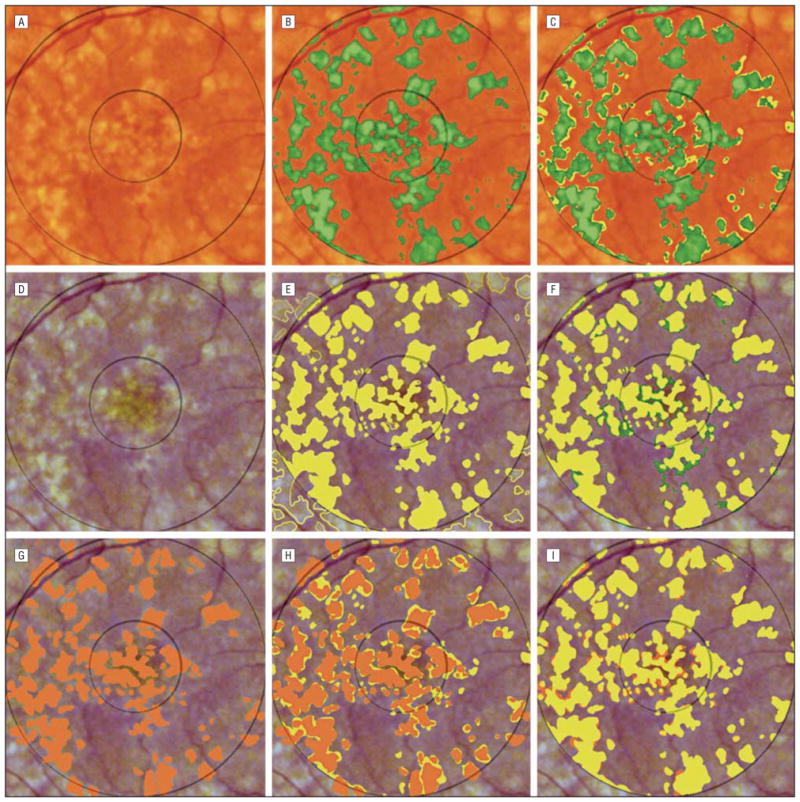
Twenty images from 17 patients were selected as representative of common manifestations of drusen. Image preprocessing included automated color balancing and, where necessary, manual segmentation of confounding lesions such as geographic atrophy (3 images). The operator then chose among 3 automated processing options suggested by predominant drusen type. Automated processing consisted of elimination of background variability by a mathematical model and subsequent histogram-based threshold selection. A retinal specialist using a graphic tablet while viewing stereo pairs constructed digital drusen drawings for each image.

MAIN OUTCOME MEASURES

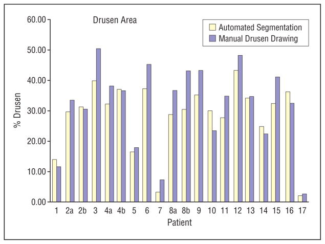
The sensitivity and specificity of drusen segmentation using the automated method with respect to manual stereoscopic drusen drawings were calculated on a rigorous pixel-by-pixel basis.

RESULTS

The median sensitivity and specificity of automated segmentation were 70% and 81%, respectively. After preprocessing and option choice, reproducibility of automated drusen segmentation was necessarily 100%.

CONCLUSIONS

Automated drusen segmentation can be reliably performed on digital fundus photographs and result in successful quantification of drusen in a more precise manner than is traditionally possible with manual stereoscopic grading of drusen. With only minor preprocessing requirements, this automated detection technique may dramatically improve our ability to monitor drusen in ARMD.

摘要

背景

年龄相关性黄斑变性(ARMD)是美国60岁以上患者视力丧失的最常见原因。在ARMD的临床评估中,观察玻璃膜疣是标志性发现。

目的

使用图像分析对ARMD患者的玻璃膜疣进行分割和量化,并比较图像分析分割与玻璃膜疣立体手工分级的效果。

设计

回顾性研究。

地点

大学转诊中心。患者照片从正在进行的哥伦比亚大学黄斑遗传学研究中已知患有ARMD的患者的可用数据库中随机选择。所有患者均为白人且年龄超过60岁。

干预措施

从17例患者中选择20张图像作为玻璃膜疣常见表现的代表。图像预处理包括自动颜色平衡,必要时对手动分割混杂病变,如地图样萎缩(3张图像)。然后,操作员从主要玻璃膜疣类型建议的3种自动处理选项中进行选择。自动处理包括通过数学模型消除背景变异性,以及随后基于直方图的阈值选择。一名视网膜专家在查看立体图像对时使用数位板为每张图像构建数字玻璃膜疣图。

主要观察指标

在严格的逐像素基础上计算使用自动方法进行玻璃膜疣分割相对于手动立体玻璃膜疣图的敏感性和特异性。

结果

自动分割的中位敏感性和特异性分别为70%和81%。经过预处理和选项选择后,自动玻璃膜疣分割的可重复性必然为100%。

结论

自动玻璃膜疣分割可以在数字眼底照片上可靠地进行,并能以比传统手动立体分级更精确的方式成功量化玻璃膜疣。由于仅需少量预处理要求,这种自动检测技术可能会显著提高我们监测ARMD中玻璃膜疣的能力。

https://cdn.ncbi.nlm.nih.gov/pmc/blobs/6fb7/2884376/a066988aaef1/nihms204457f4.jpg
https://cdn.ncbi.nlm.nih.gov/pmc/blobs/6fb7/2884376/7ce411948c8f/nihms204457f1.jpg
https://cdn.ncbi.nlm.nih.gov/pmc/blobs/6fb7/2884376/50c28e0eaf3c/nihms204457f2.jpg
https://cdn.ncbi.nlm.nih.gov/pmc/blobs/6fb7/2884376/75e974bb080e/nihms204457f3.jpg
https://cdn.ncbi.nlm.nih.gov/pmc/blobs/6fb7/2884376/a066988aaef1/nihms204457f4.jpg

相似文献

1
Automated detection of macular drusen using geometric background leveling and threshold selection.利用几何背景均衡和阈值选择自动检测黄斑玻璃膜疣。
Arch Ophthalmol. 2005 Feb;123(2):200-6. doi: 10.1001/archopht.123.2.200.
2
Automated drusen detection in retinal images using analytical modelling algorithms.基于分析建模算法的视网膜图像自动 drusen 检测
Biomed Eng Online. 2011 Jul 12;10:59. doi: 10.1186/1475-925X-10-59.
3
Automated detection of age-related macular degeneration in color fundus photography: a systematic review.彩色眼底照相中与年龄相关的黄斑变性的自动检测:系统评价。
Surv Ophthalmol. 2019 Jul-Aug;64(4):498-511. doi: 10.1016/j.survophthal.2019.02.003. Epub 2019 Feb 14.
4
Comparison of nonmydriatic digitized video fundus images with standard 35-mm slides to screen for and identify specific lesions of age-related macular degeneration.非散瞳数字化视频眼底图像与标准35毫米幻灯片用于年龄相关性黄斑变性特定病变筛查和识别的比较。
Retina. 2002 Feb;22(1):59-64. doi: 10.1097/00006982-200202000-00011.
5
Drusen regression is associated with local changes in fundus autofluorescence in intermediate age-related macular degeneration.玻璃膜疣消退与年龄相关性中间型黄斑变性的眼底自发荧光的局部变化有关。
Am J Ophthalmol. 2013 Sep;156(3):532-542.e1. doi: 10.1016/j.ajo.2013.04.031. Epub 2013 Jul 3.
6
Computer-assisted, interactive fundus image processing for macular drusen quantitation.用于黄斑玻璃膜疣定量分析的计算机辅助交互式眼底图像处理
Ophthalmology. 1999 Jun;106(6):1119-25. doi: 10.1016/S0161-6420(99)90257-9.
7
A method of drusen measurement based on reconstruction of fundus background reflectance.一种基于眼底背景反射率重建的玻璃膜疣测量方法。
Br J Ophthalmol. 2005 Jan;89(1):87-91. doi: 10.1136/bjo.2004.042937.
8
Quantitative image analysis of macular drusen from fundus photographs and scanning laser ophthalmoscope images.基于眼底照片和扫描激光眼底镜图像对黄斑玻璃膜疣进行定量图像分析。
Eye (Lond). 1995;9 ( Pt 1):48-55. doi: 10.1038/eye.1995.7.
9
A method of drusen measurement based on the geometry of fundus reflectance.一种基于眼底反射几何形状的玻璃膜疣测量方法。
Biomed Eng Online. 2003 Apr 18;2:10. doi: 10.1186/1475-925x-2-10.
10
Comparison of drusen area detected by spectral domain optical coherence tomography and color fundus imaging.比较频域光相干断层扫描和眼底彩色成像检测到的玻璃膜疣面积。
Invest Ophthalmol Vis Sci. 2013 Apr 3;54(4):2429-34. doi: 10.1167/iovs.12-11569.

引用本文的文献

1
The possibility of the combination of OCT and fundus images for improving the diagnostic accuracy of deep learning for age-related macular degeneration: a preliminary experiment.OCT 与眼底图像相结合提高深度学习诊断年龄相关性黄斑变性准确性的可能性:一项初步实验。
Med Biol Eng Comput. 2019 Mar;57(3):677-687. doi: 10.1007/s11517-018-1915-z. Epub 2018 Oct 22.
2
Automatic Screening and Grading of Age-Related Macular Degeneration from Texture Analysis of Fundus Images.基于眼底图像纹理分析的年龄相关性黄斑变性自动筛查与分级
J Ophthalmol. 2016;2016:5893601. doi: 10.1155/2016/5893601. Epub 2016 Apr 14.
3
Dynamic Drusen Remodelling in Participants of the Nutritional AMD Treatment-2 (NAT-2) Randomized Trial.

本文引用的文献

1
A method of drusen measurement based on reconstruction of fundus background reflectance.一种基于眼底背景反射率重建的玻璃膜疣测量方法。
Br J Ophthalmol. 2005 Jan;89(1):87-91. doi: 10.1136/bjo.2004.042937.
2
Current concepts in the pathogenesis of age-related macular degeneration.年龄相关性黄斑变性发病机制的当前概念
Arch Ophthalmol. 2004 Apr;122(4):598-614. doi: 10.1001/archopht.122.4.598.
3
Photographic patterns in macular images: representation by a mathematical model.黄斑图像中的摄影模式:用数学模型表示
营养性年龄相关性黄斑变性治疗-2(NAT-2)随机试验参与者中的动态玻璃膜疣重塑
PLoS One. 2016 Feb 22;11(2):e0149219. doi: 10.1371/journal.pone.0149219. eCollection 2016.
4
Fully automatic segmentation of fluorescein leakage in subjects with diabetic macular edema.糖尿病性黄斑水肿患者荧光素渗漏的全自动分割
Invest Ophthalmol Vis Sci. 2015 Jan 29;56(3):1482-92. doi: 10.1167/iovs.14-15457.
5
A survey on computer aided diagnosis for ocular diseases.一项关于眼科疾病计算机辅助诊断的调查。
BMC Med Inform Decis Mak. 2014 Aug 31;14:80. doi: 10.1186/1472-6947-14-80.
6
Automated drusen segmentation and quantification in SD-OCT images.SD-OCT 图像中的自动 drusen 分割和定量。
Med Image Anal. 2013 Dec;17(8):1058-72. doi: 10.1016/j.media.2013.06.003. Epub 2013 Jul 2.
7
Drusen regression is associated with local changes in fundus autofluorescence in intermediate age-related macular degeneration.玻璃膜疣消退与年龄相关性中间型黄斑变性的眼底自发荧光的局部变化有关。
Am J Ophthalmol. 2013 Sep;156(3):532-542.e1. doi: 10.1016/j.ajo.2013.04.031. Epub 2013 Jul 3.
8
Optimisation of an automated drusen-quantifying software for the analysis of drusen distribution in patients with age-related macular degeneration.用于分析年龄相关性黄斑变性患者中玻璃膜疣分布的自动化玻璃膜疣定量软件的优化。
Eye (Lond). 2013 Apr;27(4):554-60. doi: 10.1038/eye.2012.292. Epub 2013 Jan 11.
9
Automatic segmentation of closed-contour features in ophthalmic images using graph theory and dynamic programming.使用图论和动态规划对眼科图像中的闭合轮廓特征进行自动分割。
Biomed Opt Express. 2012 May 1;3(5):1127-40. doi: 10.1364/BOE.3.001127. Epub 2012 Apr 26.
10
Decoding simulated neurodynamics predicts the perceptual consequences of age-related macular degeneration.解码模拟神经动力学可预测年龄相关性黄斑变性的感知后果。
J Vis. 2011 Dec 5;11(14):4. doi: 10.1167/11.14.4.
J Biomed Opt. 2004 Jan-Feb;9(1):162-72. doi: 10.1117/1.1630604.
4
A method of drusen measurement based on the geometry of fundus reflectance.一种基于眼底反射几何形状的玻璃膜疣测量方法。
Biomed Eng Online. 2003 Apr 18;2:10. doi: 10.1186/1475-925x-2-10.
5
Detection and segmentation of drusen deposits on human retina: potential in the diagnosis of age-related macular degeneration.人视网膜上玻璃膜疣沉积物的检测与分割:在年龄相关性黄斑变性诊断中的潜力。
Med Image Anal. 2003 Mar;7(1):95-108. doi: 10.1016/s1361-8415(02)00093-2.
6
A new approach of geodesic reconstruction for drusen segmentation in eye fundus images.一种用于眼底图像中玻璃膜疣分割的测地线重建新方法。
IEEE Trans Med Imaging. 2001 Dec;20(12):1321-33. doi: 10.1109/42.974927.
7
Computer-assisted, interactive fundus image processing for macular drusen quantitation.用于黄斑玻璃膜疣定量分析的计算机辅助交互式眼底图像处理
Ophthalmology. 1999 Jun;106(6):1119-25. doi: 10.1016/S0161-6420(99)90257-9.
8
A pilot randomized controlled study on the effect of laser photocoagulation of confluent soft macular drusen.一项关于融合性软性黄斑玻璃疣激光光凝效果的前瞻性随机对照研究。
Ophthalmology. 1997 Apr;104(4):623-31. doi: 10.1016/s0161-6420(97)30261-9.
9
Bilateral macular drusen in age-related macular degeneration. Prognosis and risk factors.年龄相关性黄斑变性中的双侧黄斑玻璃膜疣。预后及危险因素。
Ophthalmology. 1994 Sep;101(9):1522-8. doi: 10.1016/s0161-6420(94)31139-0.
10
Automated extraction and quantification of macular drusen from fundal photographs.从眼底照片中自动提取和量化黄斑玻璃膜疣。
Aust N Z J Ophthalmol. 1994 Feb;22(1):7-12. doi: 10.1111/j.1442-9071.1994.tb01688.x.