Shechter Guy, Resar Jon R, McVeigh Elliot R
Lab of Cardiac Energetics, NHLBI, Bethesda, Maryland 20892-1061, USA.
Med Phys. 2005 Jan;32(1):255-62. doi: 10.1118/1.1836291.
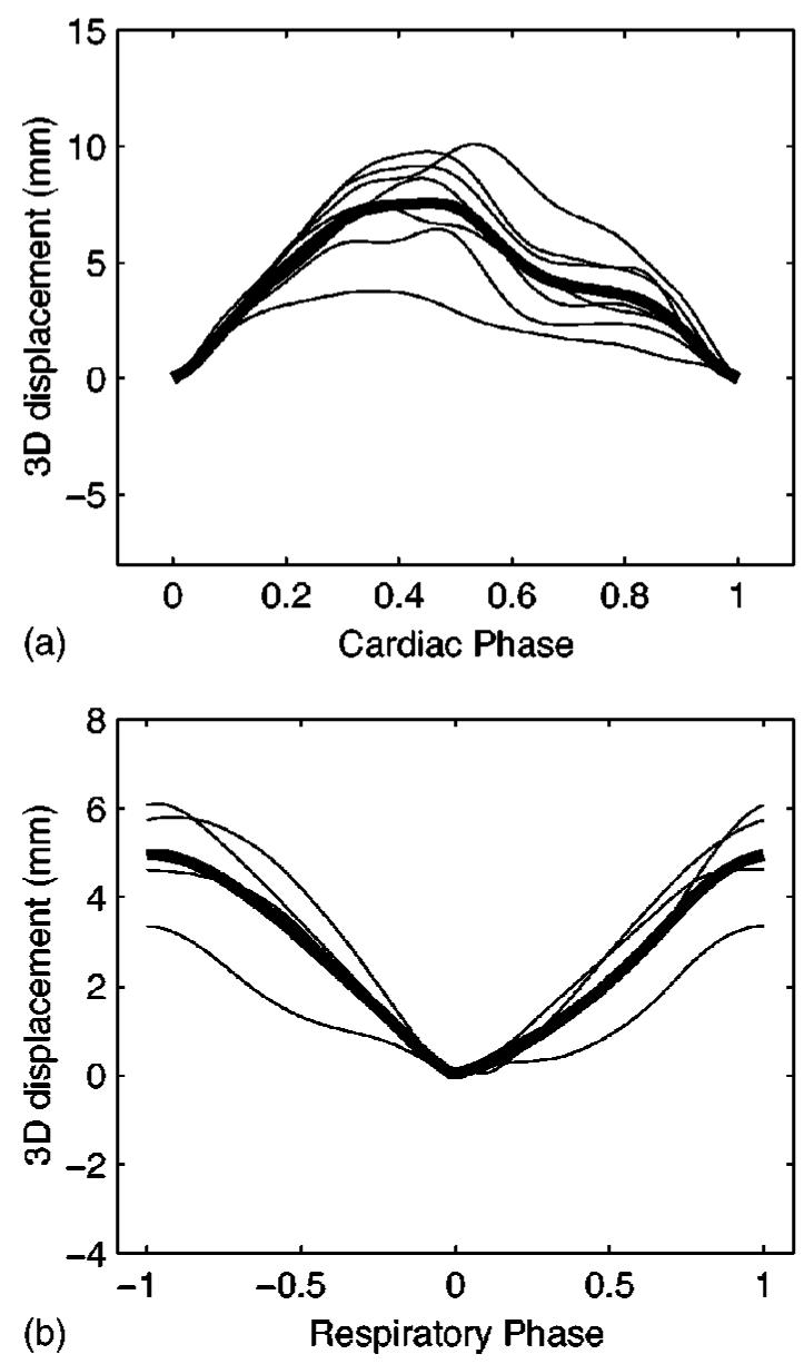
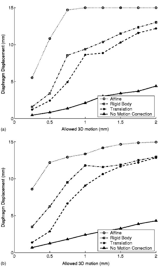
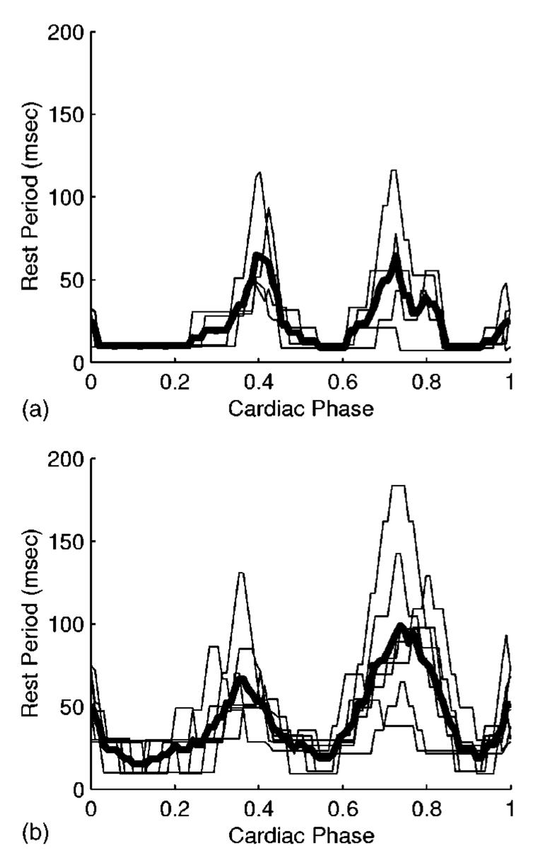
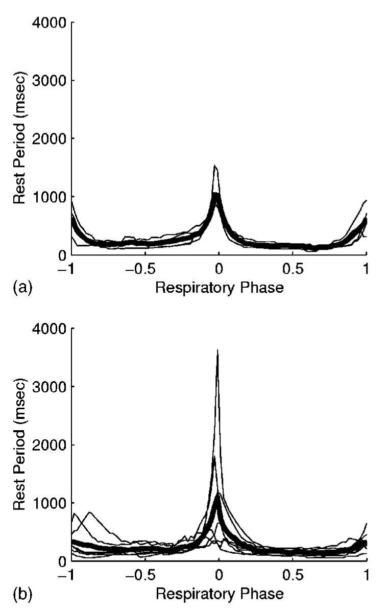
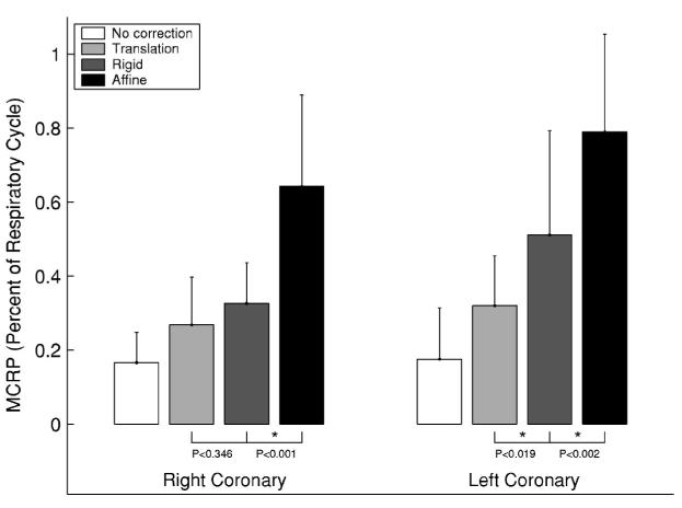
Magnetic resonance (MR) and computed tomography coronary imaging is susceptible to artifacts caused by motion of the heart. The presence of rest periods during the cardiac and respiratory cycles suggests that images free of motion artifacts could be acquired. In this paper, we studied the rest period (RP) duration of the coronary arteries during a cardiac contraction and a tidal respiratory cycle. We also studied whether three MR motion correction methods could be used to increase the respiratory RP duration. Free breathing x-ray coronary angiograms were acquired in ten patients. The three-dimensional (3D) structure of the coronary arteries was reconstructed from a biplane acquisition using stereo reconstruction methods. The 3D motion of the arterial model was then recovered using an automatic motion tracking algorithm. The motion field was then decomposed into separate cardiac and respiratory components using a cardiac respiratory parametric model. For the proximal-to-middle segments of the right coronary artery (RCA), a cardiac RP (<1 mm 3D displacement) of 76+/-34 ms was measured at end systole (ES), and 65+/-42 ms in mid-diastole (MD). The cardiac RP was 80+/-25 ms at ES and 112+/-42 ms at MD for the proximal 5 cm of the left coronary tree. At end expiration, the respiratory RP (in percent of the respiratory period) was 26+/-8% for the RCA and 27+/-17% for the left coronary tree. Left coronary respiratory RP (<0.5 mm 3D displacement) increased with translation (32% of the respiratory period), rigid body (51%), and affine (79%) motion correction. The RCA respiratory RP using translational (27%) and rigid body (33%) motion correction were not statistically different from each other. Measurements of the cardiac and respiratory rest periods will improve our understanding of the temporal and spatial resolution constraints for coronary imaging.
磁共振(MR)和计算机断层扫描冠状动脉成像容易受到心脏运动引起的伪影影响。心脏和呼吸周期中存在静止期,这表明可以获取无运动伪影的图像。在本文中,我们研究了心脏收缩和潮式呼吸周期中冠状动脉的静止期(RP)持续时间。我们还研究了三种MR运动校正方法是否可用于增加呼吸RP持续时间。对10名患者进行了自由呼吸状态下的X射线冠状动脉造影。使用立体重建方法从双平面采集重建冠状动脉的三维(3D)结构。然后使用自动运动跟踪算法恢复动脉模型的3D运动。接着使用心脏呼吸参数模型将运动场分解为单独的心脏和呼吸分量。对于右冠状动脉(RCA)的近端至中段,在收缩末期(ES)测量到心脏RP(3D位移<1 mm)为76±34 ms,在舒张中期(MD)为65±42 ms。左冠状动脉树近端5 cm处,在ES时心脏RP为80±25 ms,在MD时为112±42 ms。在呼气末,RCA的呼吸RP(占呼吸周期的百分比)为26±8%,左冠状动脉树为27±17%。左冠状动脉呼吸RP(3D位移<0.5 mm)在平移(占呼吸周期的32%)、刚体(51%)和仿射(79%)运动校正后增加。使用平移(27%)和刚体(33%)运动校正的RCA呼吸RP在统计学上彼此无差异。对心脏和呼吸静止期进行测量将有助于我们更好地理解冠状动脉成像的时间和空间分辨率限制。