Nett Brian E, Leng Shuai, Zambelli Joseph N, Reeder Scott B, Speidel Michael A, Chen Guang-Hong
Department of Medical Physics, University of Wisconsin Madison, J5/M174, Clinical Science Center, 600 Highland Avenue, Madison, WI 53792-1590, USA.
Acad Radiol. 2008 Jan;15(1):93-106. doi: 10.1016/j.acra.2007.07.003.
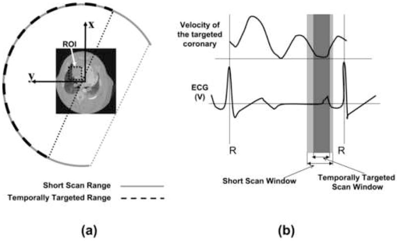
Existing cardiac imaging methods do not allow for improved temporal resolution when considering a targeted region of interest (ROI). The imaging method presented here enables improved temporal resolution for ROI imaging (namely, a reconstruction volume smaller than the complete field of view). Clinically, temporally targeted reconstruction would not change the primary means of reconstructing and evaluating images, but rather would enable the adjunct technique of ROI imaging, with improved temporal resolution compared with standard reconstruction ( approximately 20% smaller temporal scan window). In gated cardiac computed tomography (CT) scans improved temporal resolution directly translates into a reduction in motion artifacts for rapidly moving objects such as the coronary arteries.
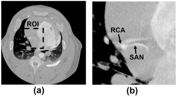
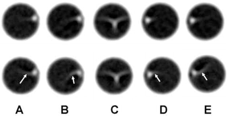
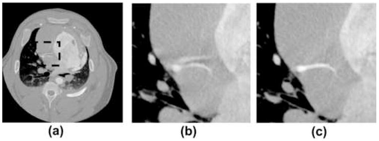
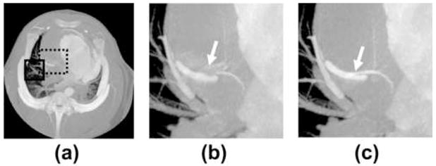
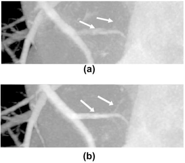
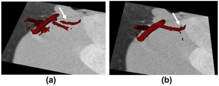
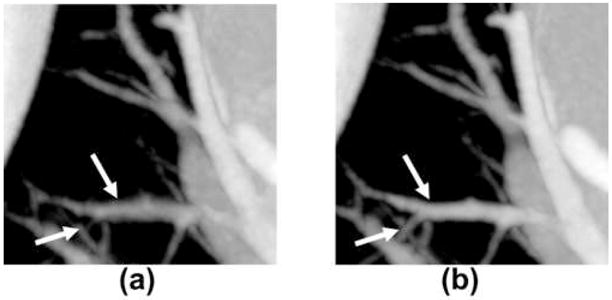
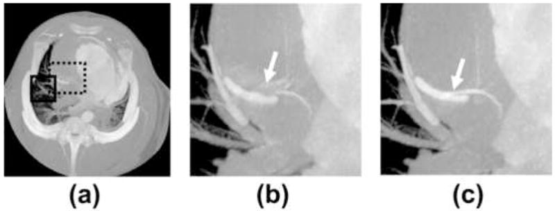
Retrospectively electrocardiogram gated coronary angiography data from a 64-slice CT system were used. A motion phantom simulating the motion profile of a coronary artery was constructed and scanned. Additionally, an in vivo study was performed using a porcine model. Comparisons between the new reconstruction technique and the standard reconstruction are given for an ROI centered on the right coronary artery, and a pulmonary ROI.
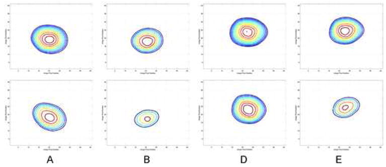
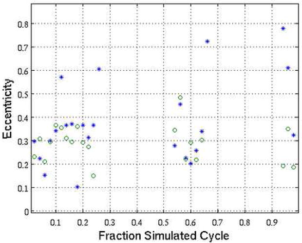
In both a well-controlled motion model and a porcine model results show a decrease in motion induced artifacts including motion blur and streak artifacts from contrast enhanced vessels within the targeted ROIs, as assessed through both qualitative and quantitative observations.
Temporally targeted reconstruction techniques demonstrate the potential to reduce motion artifacts in coronary CT. Further study is warranted to demonstrate the conditions under which this technique will offer direct clinical utility. Improvement in temporal resolution for gated cardiac scans has implications for improving: contrast enhanced CT angiography, calcium scoring, and assessment of the pulmonary anatomy.
现有的心脏成像方法在考虑目标感兴趣区域(ROI)时,无法提高时间分辨率。本文介绍的成像方法能够提高ROI成像的时间分辨率(即,重建体积小于完整视野)。在临床上,时间靶向重建不会改变图像重建和评估的主要方式,而是能够实现ROI成像的辅助技术,与标准重建相比,时间分辨率得到提高(时间扫描窗口缩小约20%)。在门控心脏计算机断层扫描(CT)中,提高时间分辨率直接转化为减少快速移动物体(如冠状动脉)的运动伪影。
使用来自64层CT系统的回顾性心电图门控冠状动脉造影数据。构建并扫描了一个模拟冠状动脉运动轮廓的运动模型。此外,使用猪模型进行了一项体内研究。给出了以右冠状动脉为中心的ROI和肺部ROI的新重建技术与标准重建之间的比较。
在良好控制的运动模型和猪模型中,结果均显示运动诱导伪影减少,包括通过定性和定量观察评估的目标ROI内对比增强血管的运动模糊和条纹伪影。
时间靶向重建技术显示出减少冠状动脉CT运动伪影的潜力。有必要进行进一步研究以证明该技术具有直接临床应用价值的条件。门控心脏扫描时间分辨率的提高对改善以下方面具有重要意义:对比增强CT血管造影、钙化评分以及肺部解剖结构评估。