Wood Bradford J, Zhang Hui, Durrani Amir, Glossop Neil, Ranjan Sohan, Lindisch David, Levy Eliott, Banovac Filip, Borgert Joern, Krueger Sascha, Kruecker Jochen, Viswanathan Anand, Cleary Kevin
Diagnostic Radiology Department, National Institutes of Health Clinical Center, Building 10, Room 1C-660, Bethesda, Maryland 20892, USA.
J Vasc Interv Radiol. 2005 Apr;16(4):493-505. doi: 10.1097/01.RVI.0000148827.62296.B4.
To assess the feasibility of the use of preprocedural imaging for guide wire, catheter, and needle navigation with electromagnetic tracking in phantom and animal models.
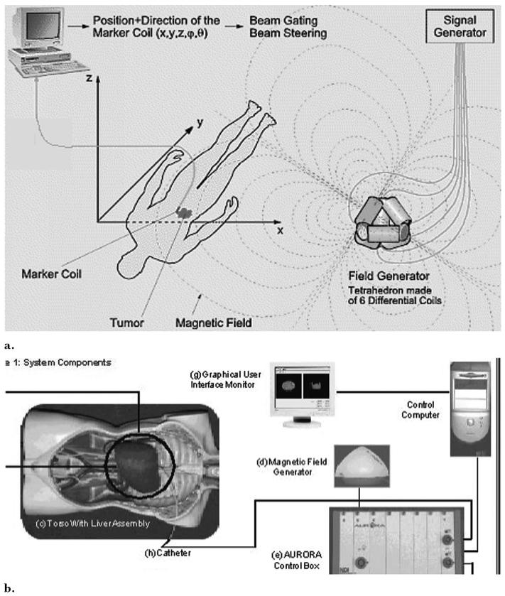
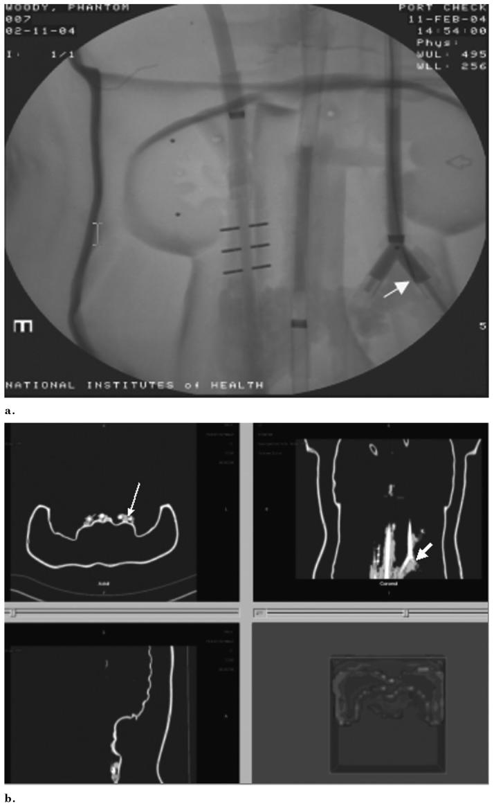
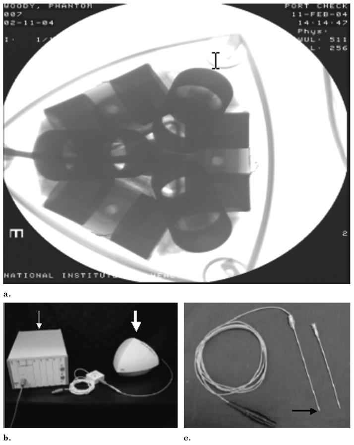
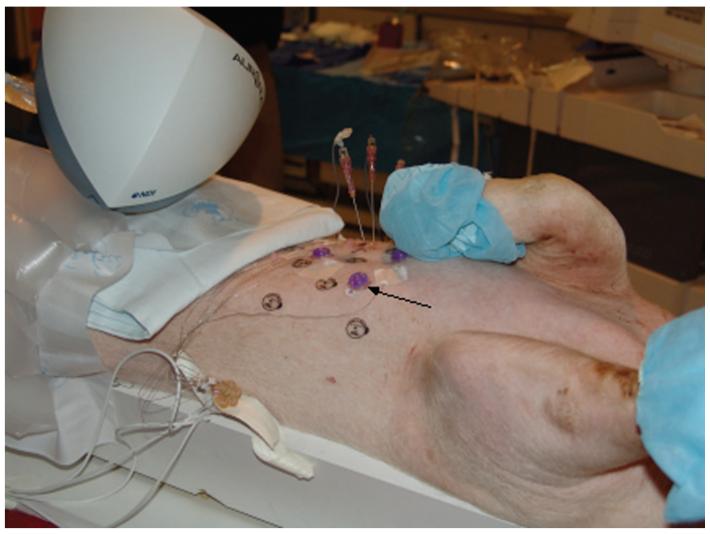
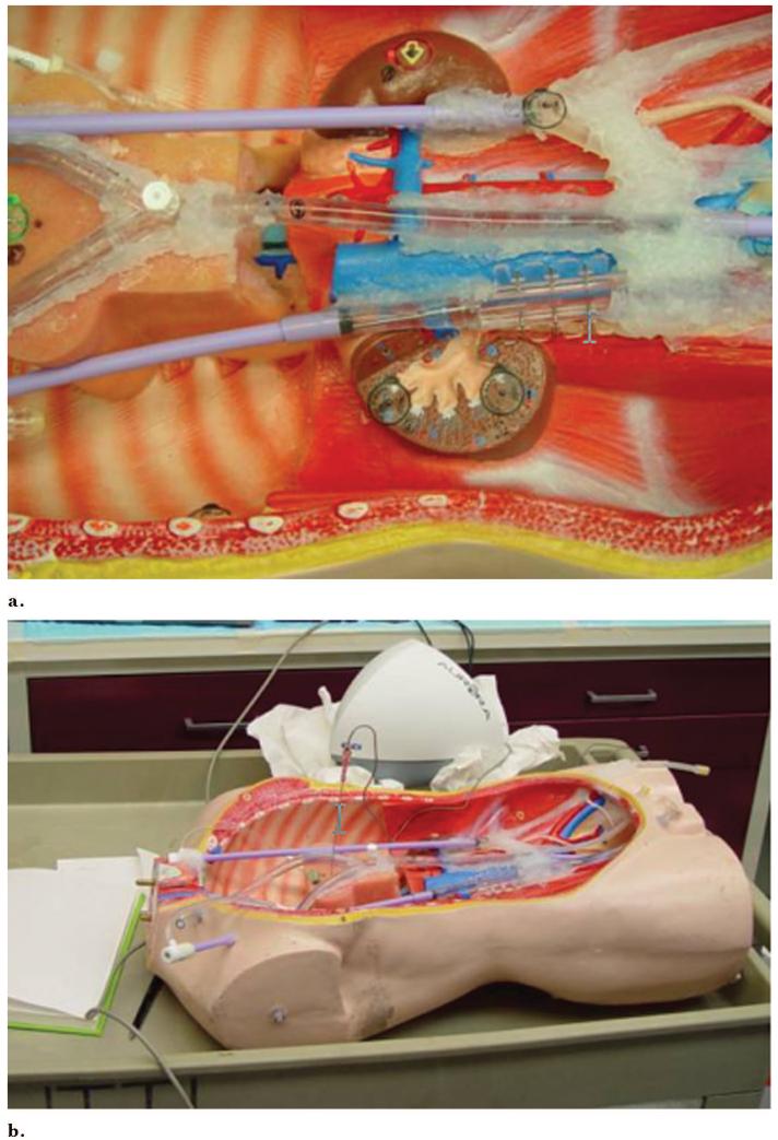
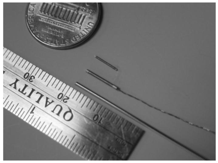
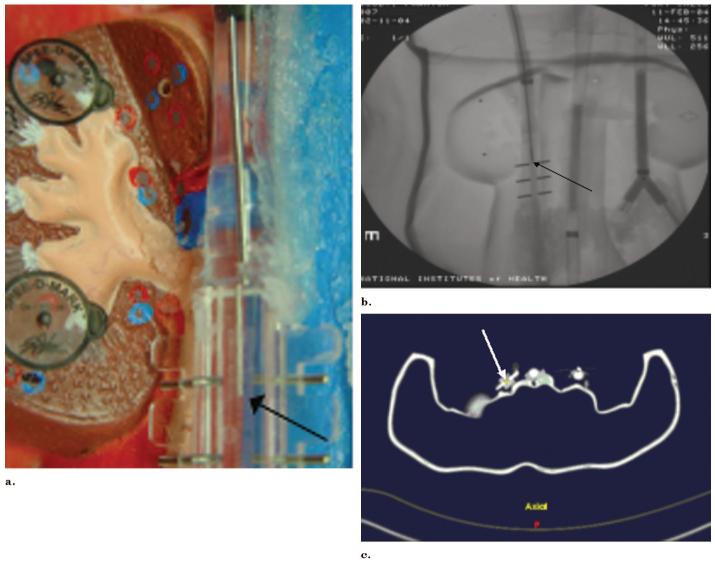
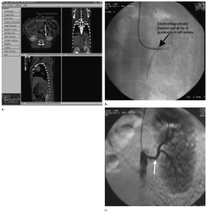
An image-guided intervention software system was developed based on open-source software components. Catheters, needles, and guide wires were constructed with small position and orientation sensors in the tips. A tetrahedral-shaped weak electromagnetic field generator was placed in proximity to an abdominal vascular phantom or three pigs on the angiography table. Preprocedural computed tomographic (CT) images of the phantom or pig were loaded into custom-developed tracking, registration, navigation, and rendering software. Devices were manipulated within the phantom or pig with guidance from the previously acquired CT scan and simultaneous real-time angiography. Navigation within positron emission tomography (PET) and magnetic resonance (MR) volumetric datasets was also performed. External and endovascular fiducials were used for registration in the phantom, and registration error and tracking error were estimated.
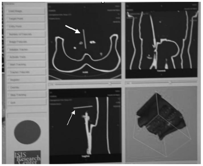
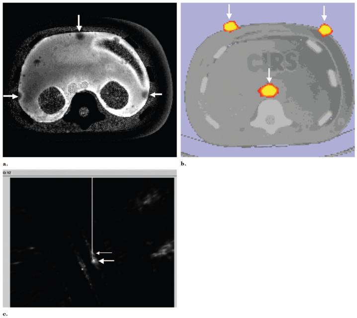
The CT scan position of the devices within phantoms and pigs was accurately determined during angiography and biopsy procedures, with manageable error for some applications. Preprocedural CT depicted the anatomy in the region of the devices with real-time position updating and minimal registration error and tracking error (<5 mm). PET can also be used with this system to guide percutaneous biopsies to the most metabolically active region of a tumor.
Previously acquired CT, MR, or PET data can be accurately codisplayed during procedures with reconstructed imaging based on the position and orientation of catheters, guide wires, or needles. Multimodality interventions are feasible by allowing the real-time updated display of previously acquired functional or morphologic imaging during angiography, biopsy, and ablation.
评估在体模和动物模型中,使用术前成像结合电磁跟踪技术来引导导丝、导管和穿刺针的可行性。
基于开源软件组件开发了一种图像引导介入软件系统。导管、穿刺针和导丝的尖端装有小型位置和方向传感器。在血管造影台上,将一个四面体形状的弱电磁场发生器放置在腹部血管体模或三头猪附近。将体模或猪的术前计算机断层扫描(CT)图像加载到定制开发的跟踪、配准、导航和渲染软件中。在先前获取的CT扫描和同步实时血管造影的引导下,在体模或猪体内操作设备。还在正电子发射断层扫描(PET)和磁共振(MR)容积数据集中进行导航。使用外部和血管内基准标记在体模中进行配准,并估计配准误差。
在血管造影和活检过程中,能够准确确定体模和猪体内设备的CT扫描位置,对于某些应用而言,误差在可控制范围内。术前CT能够实时更新位置,以最小的配准误差和跟踪误差(<5毫米)描绘设备所在区域的解剖结构。PET也可用于该系统,以引导经皮活检至肿瘤代谢最活跃的区域。
在手术过程中,根据导管、导丝或穿刺针的位置和方向,通过重建成像可以准确地共同显示先前获取的CT、MR或PET数据。通过在血管造影、活检和消融过程中实时更新显示先前获取的功能或形态学成像,多模态干预是可行的。