Hattori Koji, Takakura Yoshinori, Ohgushi Hajime, Habata Takashi, Uematsu Kota, Yamauchi Jun, Yamashita Kenji, Fukuchi Takashi, Sato Masao, Ikeuchi Ken
Department of Orthopaedic Surgery, Nara Medical University, Nara, Japan.
Arthritis Res Ther. 2005;7(3):R552-9. doi: 10.1186/ar1710. Epub 2005 Mar 1.
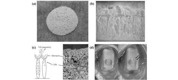
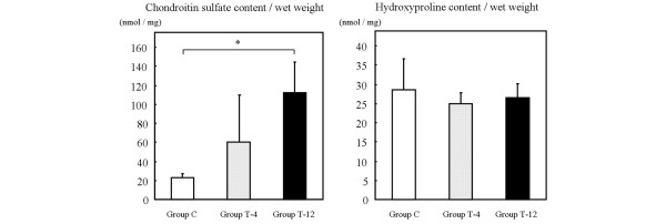
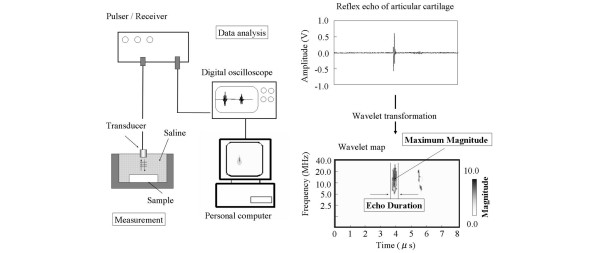
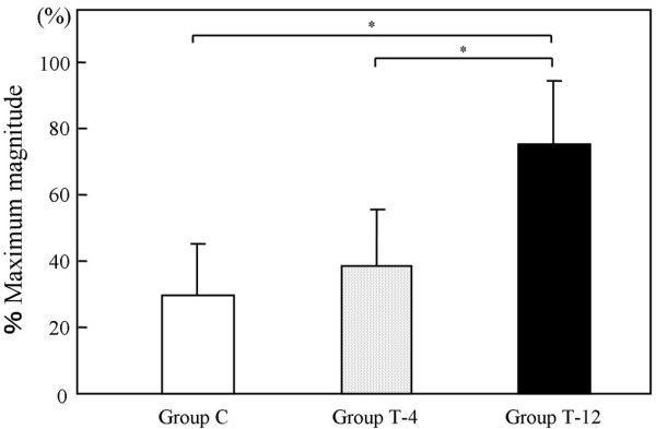
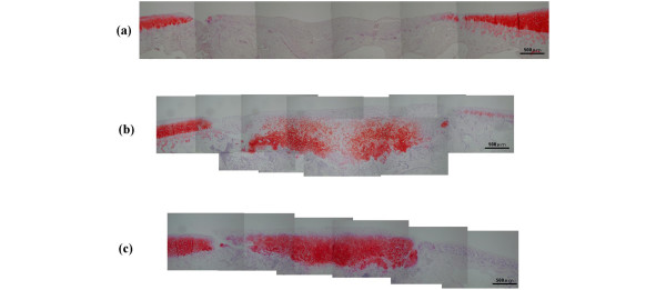
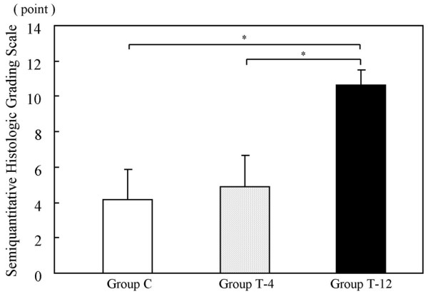
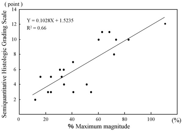
Articular cartilage (hyaline cartilage) defects resulting from traumatic injury or degenerative joint disease do not repair themselves spontaneously. Therefore, such defects may require novel regenerative strategies to restore biologically and biomechanically functional tissue. Recently, tissue engineering using a complex of cells and scaffold has emerged as a new approach for repairing cartilage defects and restoring cartilage function. With the advent of this new technology, accurate methods for evaluating articular cartilage have become important. In particular, in vivo evaluation is essential for determining the best treatment. However, without a biopsy, which causes damage, articular cartilage cannot be accurately evaluated in a clinical context. We have developed a novel system for evaluating articular cartilage, in which the acoustic properties of the cartilage are measured by introducing an ultrasonic probe during arthroscopy of the knee joint. The purpose of the current study was to determine the efficacy of this ultrasound system for evaluating tissue-engineered cartilage in an experimental model involving implantation of a cell/scaffold complex into rabbit knee joint defects. Ultrasonic echoes from the articular cartilage were converted into a wavelet map by wavelet transformation. On the wavelet map, the percentage maximum magnitude (the maximum magnitude of the measurement area of the operated knee divided by that of the intact cartilage of the opposite, nonoperated knee; %MM) was used as a quantitative index of cartilage regeneration. Using this index, the tissue-engineered cartilage was examined to elucidate the relations between ultrasonic analysis and biochemical and histological analyses. The %MM increased over the time course of the implant and all the hyaline-like cartilage samples from the histological findings had a high %MM. Correlations were observed between the %MM and the semiquantitative histologic grading scale scores from the histological findings. In the biochemical findings, the chondroitin sulfate content increased over the time course of the implant, whereas the hydroxyproline content remained constant. The chondroitin sulfate content showed a similarity to the results of the %MM values. Ultrasonic measurements were found to predict the regeneration process of the tissue-engineered cartilage as a minimally invasive method. Therefore, ultrasonic evaluation using a wavelet map can support the evaluation of tissue-engineered cartilage using cell/scaffold complexes.
创伤性损伤或退行性关节疾病导致的关节软骨(透明软骨)缺损无法自行修复。因此,此类缺损可能需要新的再生策略来恢复具有生物学和生物力学功能的组织。近年来,利用细胞与支架复合物的组织工程学已成为修复软骨缺损和恢复软骨功能的新方法。随着这项新技术的出现,准确评估关节软骨的方法变得至关重要。特别是,体内评估对于确定最佳治疗方案必不可少。然而,在临床环境中,若不进行会造成损伤的活检,就无法准确评估关节软骨。我们开发了一种评估关节软骨的新系统,该系统通过在膝关节关节镜检查期间引入超声探头来测量软骨的声学特性。本研究的目的是在将细胞/支架复合物植入兔膝关节缺损的实验模型中,确定这种超声系统评估组织工程软骨的效果。关节软骨的超声回波通过小波变换转换为小波图。在小波图上,最大幅度百分比(手术膝关节测量区域的最大幅度除以对侧未手术膝关节完整软骨的最大幅度;%MM)被用作软骨再生的定量指标。利用该指标,对组织工程软骨进行检查,以阐明超声分析与生化分析和组织学分析之间的关系。%MM在植入后的时间进程中增加,并且组织学结果显示所有类透明软骨样本的%MM都很高。在%MM与组织学结果的半定量组织学分级量表评分之间观察到相关性。在生化结果中,硫酸软骨素含量在植入后的时间进程中增加,而羟脯氨酸含量保持不变。硫酸软骨素含量与%MM值的结果相似。超声测量被发现作为一种微创方法可以预测组织工程软骨的再生过程。因此,使用小波图的超声评估可以辅助对使用细胞/支架复合物的组织工程软骨进行评估。