Kim Jung Han, Jung Joo Young, Park Young lee, Hwang Sang Ik, Jung Chull Sung, Lee Sang Hak, Yoo Chong Woo
Department of Intemal Medicine, College of Medicine, Hallym University, Seoul, Korea.
Korean J Intern Med. 2005 Mar;20(1):86-9. doi: 10.3904/kjim.2005.20.1.86.
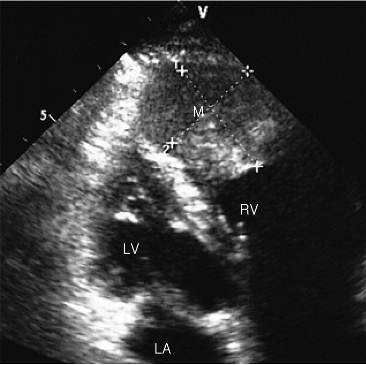
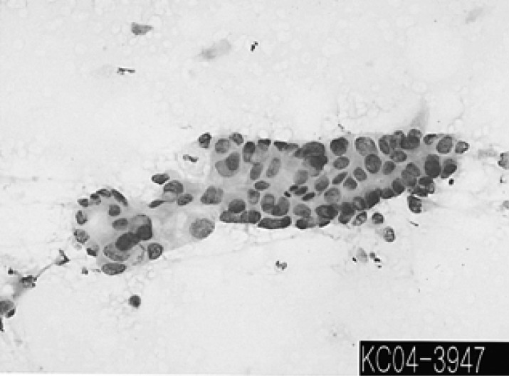
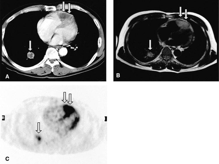
Intracardiac metastasis as the initial presentation of malignant neoplasm is very rare. We report here on a 64-year-old man with non-small cell lung cancer (NSCLC) initially presenting with intracardiac metastasis which was identified with 18-F fluorodeoxyglucose positron emission tomography (FDG PET). The patient was admitted with complaints of exertional dyspnea and vague chest discomfort that had developed a few weeks ago. Two-dimensional echocardiography revealed a heart mass attached to its akinetic wall in the right ventricular chamber. CT and MRI demonstrated a large tumor involving the epicardium and myocardium in the right ventricle, and there was a mass in the right lower lobe of the lung along with multiple lymphadenopathies. Cytologic examination of the percutaneous needle aspiration of a lymph node in the anterior mediastinum revealed malignant epithelial cell nests, and this was strongly suggestive of squamous cell carcinoma. Subsequent FDG PET confirmed that the intracardiac mass had an abnormally increased FDG uptake, and again this was strongly suggestive of malignancy. By systemically considering these imaging studies, we were able to diagnose the mass as intracardiac metastasis of NSCLC.
以心脏内转移作为恶性肿瘤的首发表现非常罕见。我们在此报告一名64岁男性,患有非小细胞肺癌(NSCLC),最初表现为心脏内转移,通过18-F氟脱氧葡萄糖正电子发射断层扫描(FDG PET)得以确诊。该患者因数周前出现的劳力性呼吸困难和模糊的胸部不适入院。二维超声心动图显示右心室腔内有一附着于运动减弱壁的心脏肿物。CT和MRI显示右心室有一个累及心外膜和心肌的大肿瘤,右肺下叶有一个肿物以及多处淋巴结肿大。经皮穿刺抽吸前纵隔淋巴结的细胞学检查发现恶性上皮细胞巢,强烈提示为鳞状细胞癌。随后的FDG PET证实心脏内肿物的FDG摄取异常增加,再次强烈提示为恶性。通过系统地综合这些影像学检查结果,我们得以将该肿物诊断为NSCLC的心脏内转移。