Wittmann Torsten, Waterman-Storer Clare M
Department of Cell Biology, The Scripps Research Institute, La Jolla, CA 92037, USA.
J Cell Biol. 2005 Jun 20;169(6):929-39. doi: 10.1083/jcb.200412114. Epub 2005 Jun 13.
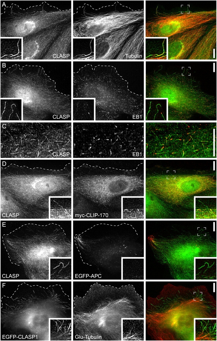
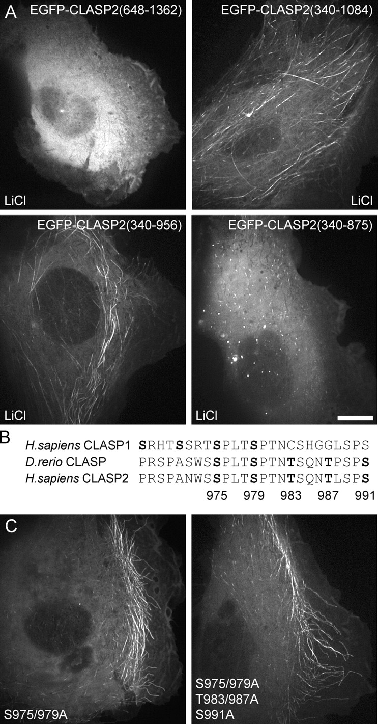
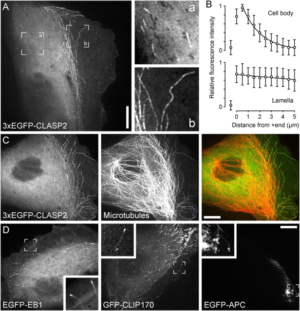
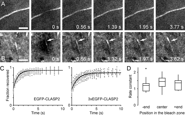
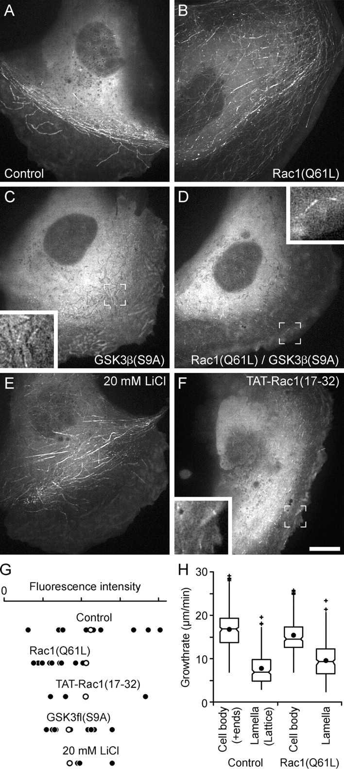
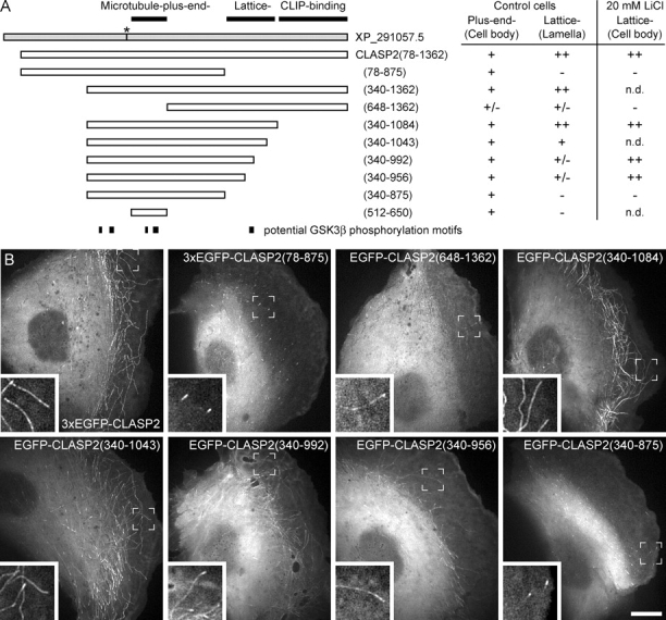
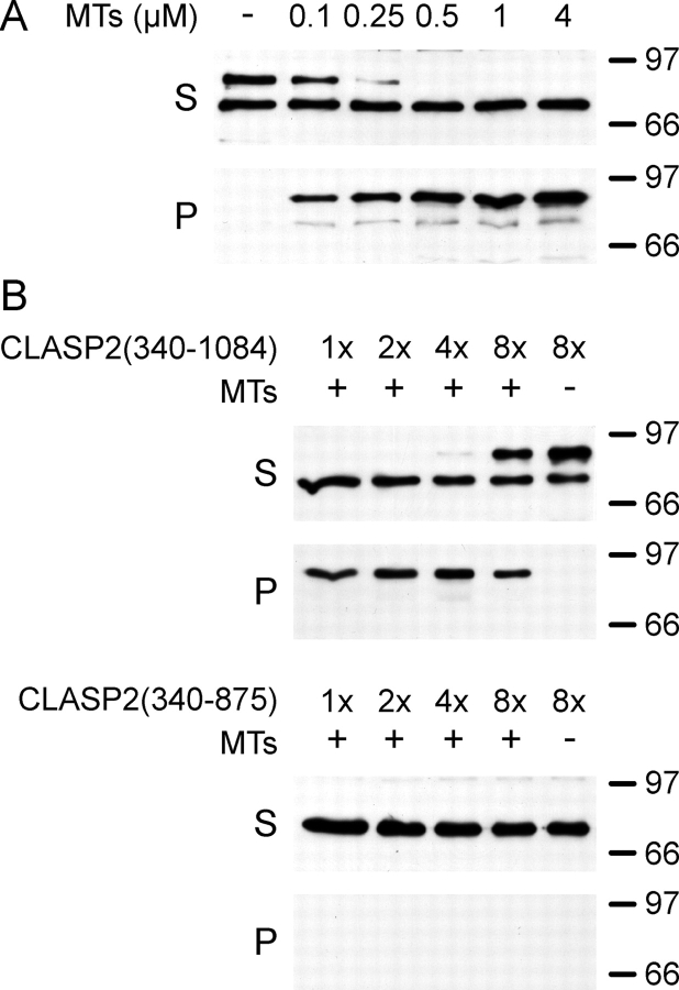
Proteins that in cells specifically bind to growing microtubule plus ends (+TIPs) are thought to play important roles in polarization of the cytoskeleton. However, most +TIPs do not show a bias of their microtubule-binding behavior toward different subcellular regions. Here, we examine the dynamics of the +TIP CLASP in migrating PtK1 epithelial cells. We find that, although CLASPs track microtubule plus ends in the cell body, they dynamically decorate the entire microtubule lattice in the leading edge lamella and lamellipodium. Microtubule lattice binding is mediated by the COOH-terminal region of the CLASP microtubule-binding domain and is regulated downstream of Rac1. Phosphorylation of sites in the NH2-terminal part of the microtubule-binding domain by glycogen synthase kinase 3beta likely regulates the affinity of CLASPs for microtubule lattices. These results demonstrate the striking difference of the microtubule cytoskeleton in the lamella as compared with the cell body and provide the first direct observation of subcellular regulation of a microtubule-associated protein in migrating cells.
细胞中特异性结合生长微管正端的蛋白质(+TIPs)被认为在细胞骨架极化中发挥重要作用。然而,大多数+TIPs在不同亚细胞区域的微管结合行为上没有表现出偏向性。在这里,我们研究了迁移的PtK1上皮细胞中+TIP CLASP的动力学。我们发现,尽管CLASP在细胞体中追踪微管正端,但它们在前缘片状伪足和片状伪足中动态修饰整个微管晶格。微管晶格结合由CLASP微管结合结构域的COOH末端区域介导,并在Rac1下游受到调节。糖原合酶激酶3β对微管结合结构域NH2末端部分位点的磷酸化可能调节CLASP对微管晶格的亲和力。这些结果表明,与细胞体相比,片状伪足中的微管细胞骨架存在显著差异,并首次直接观察到迁移细胞中微管相关蛋白的亚细胞调节。