• 文献检索
  • 文档翻译
  • 深度研究
  • 学术资讯
  • Suppr Zotero 插件Zotero 插件
  • 邀请有礼
  • 套餐&价格
  • 历史记录
应用&插件
Suppr Zotero 插件Zotero 插件浏览器插件Mac 客户端Windows 客户端微信小程序
定价
高级版会员购买积分包购买API积分包
服务
文献检索文档翻译深度研究API 文档MCP 服务
关于我们
关于 Suppr公司介绍联系我们用户协议隐私条款
关注我们

Suppr 超能文献

核心技术专利:CN118964589B侵权必究
粤ICP备2023148730 号-1Suppr @ 2026

文献检索

告别复杂PubMed语法,用中文像聊天一样搜索,搜遍4000万医学文献。AI智能推荐,让科研检索更轻松。

立即免费搜索

文件翻译

保留排版,准确专业,支持PDF/Word/PPT等文件格式,支持 12+语言互译。

免费翻译文档

深度研究

AI帮你快速写综述,25分钟生成高质量综述,智能提取关键信息,辅助科研写作。

立即免费体验

Rac1下游前沿微管和肌动蛋白动力学的调控

Regulation of leading edge microtubule and actin dynamics downstream of Rac1.

作者信息

Wittmann Torsten, Bokoch Gary M, Waterman-Storer Clare M

机构信息

Department of Cell Biology and Institute for Childhood and Neglected Diseases, La Jolla, CA 92037, USA.

出版信息

J Cell Biol. 2003 Jun 9;161(5):845-51. doi: 10.1083/jcb.200303082.

DOI:10.1083/jcb.200303082
PMID:12796474
原文链接:https://pmc.ncbi.nlm.nih.gov/articles/PMC2172968/
Abstract

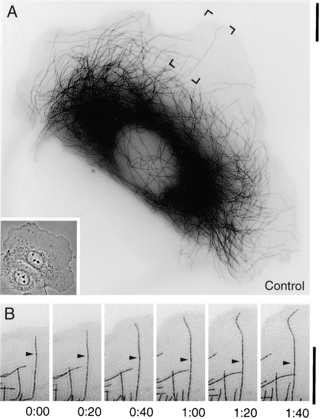
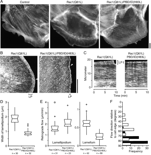
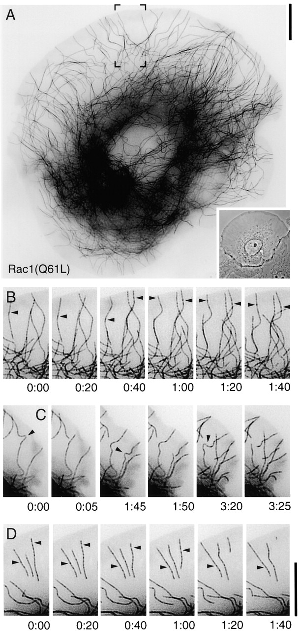
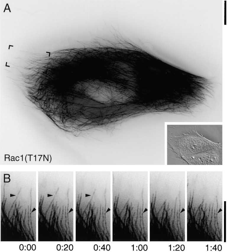
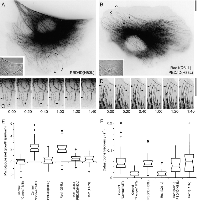
Actin in migrating cells is regulated by Rho GTPases. However, Rho proteins might also affect microtubules (MTs). Here, we used time-lapse microscopy of PtK1 cells to examine MT regulation downstream of Rac1. In these cells, "pioneer" MTs growing into leading-edge protrusions exhibited a decreased catastrophe frequency and an increased time in growth as compared with MTs further from the leading edge. Constitutively active Rac1(Q61L) promoted pioneer behavior in most MTs, whereas dominant-negative Rac1(T17N) eliminated pioneer MTs, indicating that Rac1 is a regulator of MT dynamics in vivo. Rac1(Q61L) also enhanced MT turnover through stimulation of MT retrograde flow and breakage. Inhibition of p21-activated kinases (Paks), downstream effectors of Rac1, inhibited Rac1(Q61L)-induced MT growth and retrograde flow. In addition, Rac1(Q61L) promoted lamellipodial actin polymerization and Pak-dependent retrograde flow. Together, these results indicate coordinated regulation of the two cytoskeletal systems in the leading edge of migrating cells.

摘要

迁移细胞中的肌动蛋白受Rho GTP酶调节。然而,Rho蛋白也可能影响微管(MTs)。在这里,我们使用PtK1细胞的延时显微镜来检查Rac1下游的微管调节。在这些细胞中,与远离前沿的微管相比,生长到前沿突起中的“先锋”微管表现出较低的灾难频率和较长的生长时间。组成型活性Rac1(Q61L)在大多数微管中促进先锋行为,而显性负性Rac1(T17N)消除先锋微管,表明Rac1是体内微管动力学的调节因子。Rac1(Q61L)还通过刺激微管逆行流动和断裂来增强微管周转。抑制Rac1的下游效应器p21激活激酶(Paks)可抑制Rac1(Q61L)诱导的微管生长和逆行流动。此外,Rac1(Q61L)促进片状伪足肌动蛋白聚合和依赖Pak的逆行流动。总之,这些结果表明迁移细胞前沿的两个细胞骨架系统存在协同调节。

https://cdn.ncbi.nlm.nih.gov/pmc/blobs/2a37/2172968/1d739a24d760/200303082f5.jpg
https://cdn.ncbi.nlm.nih.gov/pmc/blobs/2a37/2172968/05b9b00d9887/200303082f1.jpg
https://cdn.ncbi.nlm.nih.gov/pmc/blobs/2a37/2172968/3fc3b7c6cc89/200303082f2.jpg
https://cdn.ncbi.nlm.nih.gov/pmc/blobs/2a37/2172968/e7ed4f7d50ab/200303082f3.jpg
https://cdn.ncbi.nlm.nih.gov/pmc/blobs/2a37/2172968/8aaaa1f8b7b2/200303082f4.jpg
https://cdn.ncbi.nlm.nih.gov/pmc/blobs/2a37/2172968/1d739a24d760/200303082f5.jpg

相似文献

1
Regulation of leading edge microtubule and actin dynamics downstream of Rac1.Rac1下游前沿微管和肌动蛋白动力学的调控
J Cell Biol. 2003 Jun 9;161(5):845-51. doi: 10.1083/jcb.200303082.
2
Automated screening of microtubule growth dynamics identifies MARK2 as a regulator of leading edge microtubules downstream of Rac1 in migrating cells.自动化的微管生长动态筛选鉴定 MARK2 是 Rac1 下游迁移细胞前缘微管的调节因子。
PLoS One. 2012;7(7):e41413. doi: 10.1371/journal.pone.0041413. Epub 2012 Jul 24.
3
Regulation of microtubule destabilizing activity of Op18/stathmin downstream of Rac1.Rac1下游的Op18/微管相关蛋白对微管去稳定活性的调节
J Biol Chem. 2004 Feb 13;279(7):6196-203. doi: 10.1074/jbc.M307261200. Epub 2003 Nov 26.
4
The PAK system links Rho GTPase signaling to thrombin-mediated platelet activation.PAK 系统将 Rho GTPase 信号与凝血酶介导的血小板激活联系起来。
Am J Physiol Cell Physiol. 2013 Sep;305(5):C519-28. doi: 10.1152/ajpcell.00418.2012. Epub 2013 Jun 19.
5
Actomyosin-based retrograde flow of microtubules in the lamella of migrating epithelial cells influences microtubule dynamic instability and turnover and is associated with microtubule breakage and treadmilling.迁移上皮细胞片层中基于肌动球蛋白的微管逆行流动影响微管动态不稳定性和周转,并与微管断裂和踏车行为相关。
J Cell Biol. 1997 Oct 20;139(2):417-34. doi: 10.1083/jcb.139.2.417.
6
How to grab a microtubule on the move.如何在微管移动时抓住它。
Dev Cell. 2002 Jul;3(1):2-4. doi: 10.1016/s1534-5807(02)00209-5.
7
Glioblastoma motility occurs in the absence of actin polymer.胶质母细胞瘤的迁移发生在没有肌动蛋白聚合的情况下。
Mol Biol Cell. 2011 Jul 1;22(13):2212-20. doi: 10.1091/mbc.E10-10-0849. Epub 2011 May 5.
8
Spatial regulation of CLASP affinity for microtubules by Rac1 and GSK3beta in migrating epithelial cells.迁移上皮细胞中Rac1和GSK3β对CLASP与微管亲和力的空间调控
J Cell Biol. 2005 Jun 20;169(6):929-39. doi: 10.1083/jcb.200412114. Epub 2005 Jun 13.
9
DEF6, a novel PH-DH-like domain protein, is an upstream activator of the Rho GTPases Rac1, Cdc42, and RhoA.DEF6是一种新型的类PH-DH结构域蛋白,是Rho GTP酶Rac1、Cdc42和RhoA的上游激活剂。
Exp Cell Res. 2004 Apr 1;294(2):335-44. doi: 10.1016/j.yexcr.2003.12.004.
10
A novel p21-activated kinase binds the actin and microtubule networks and induces microtubule stabilization.一种新型的p21激活激酶与肌动蛋白和微管网络结合并诱导微管稳定。
J Cell Biol. 2001 Dec 10;155(6):1029-42. doi: 10.1083/jcb.200104123.

引用本文的文献

1
Morphological regulation of wound repair astrocytes by leucine zipper-bearing kinase-AKT signaling after spinal cord injury.脊髓损伤后亮氨酸拉链激酶-AKT信号通路对创伤修复星形胶质细胞的形态学调控
Exp Neurol. 2025 Nov;393:115379. doi: 10.1016/j.expneurol.2025.115379. Epub 2025 Jul 16.
2
The Ragulator complex and lysosomal calcium release are crucial for cell migration.Ragulator复合体和溶酶体钙释放对细胞迁移至关重要。
Life Sci Alliance. 2025 Jun 10;8(8). doi: 10.26508/lsa.202403015. Print 2025 Aug.
3
Integration of focal adhesion morphogenesis and polarity by DOCK5 promotes YAP/TAZ-driven drug resistance in TNBC.

本文引用的文献

1
Fluorescent speckle microscopy (FSM) of microtubules and actin in living cells.活细胞中微管和肌动蛋白的荧光斑点显微镜检查(FSM)。
Curr Protoc Cell Biol. 2002 Feb;Chapter 4:Unit 4.10. doi: 10.1002/0471143030.cb0410s13.
2
Biology of the p21-activated kinases.p21激活激酶的生物学特性
Annu Rev Biochem. 2003;72:743-81. doi: 10.1146/annurev.biochem.72.121801.161742. Epub 2003 Mar 27.
3
A high-speed multispectral spinning-disk confocal microscope system for fluorescent speckle microscopy of living cells.一种用于活细胞荧光斑点显微镜检查的高速多光谱旋转盘共聚焦显微镜系统。
DOCK5通过整合粘着斑形态发生和极性促进三阴性乳腺癌中YAP/TAZ驱动的耐药性。
Mol Omics. 2025 May 12. doi: 10.1039/d4mo00154k.
4
How does the tubulin code facilitate directed cell migration?微管蛋白编码如何促进细胞定向迁移?
Biochem Soc Trans. 2025 Feb 21;53(1):BST20240841. doi: 10.1042/BST20240841.
5
Doublecortin reinforces microtubules to promote growth cone advance in soft environments.双皮质素加强微管,以促进生长锥在柔软环境中的前进。
Curr Biol. 2024 Dec 16;34(24):5822-5832.e5. doi: 10.1016/j.cub.2024.10.063. Epub 2024 Dec 2.
6
Characterization of the small Arabidopsis thaliana GTPase and ADP-ribosylation factor-like 2 protein TITAN 5.拟南芥小 GTP 酶和 ADP-ribosylation factor-like 2 蛋白 TITAN 5 的特性研究。
J Cell Sci. 2024 Aug 1;137(15). doi: 10.1242/jcs.262315. Epub 2024 Aug 14.
7
Spatiotemporal Coordination of Rac1 and Cdc42 at the Whole Cell Level during Cell Ruffling.细胞皱襞过程中 Rac1 和 Cdc42 在全细胞水平上的时空协调
Cells. 2023 Jun 15;12(12):1638. doi: 10.3390/cells12121638.
8
Characterization of the small GTPase and ADP-ribosylation factor-like 2 protein TITAN 5.小GTP酶和ADP核糖基化因子样2蛋白TITAN 5的特性分析
bioRxiv. 2024 May 27:2023.04.27.538563. doi: 10.1101/2023.04.27.538563.
9
Liver Proteome Alterations in Red Deer () Infected by the Giant Liver Fluke .感染大片吸虫的马鹿肝脏蛋白质组变化
Pathogens. 2022 Dec 8;11(12):1503. doi: 10.3390/pathogens11121503.
10
Septins guide noncentrosomal microtubules to promote focal adhesion disassembly in migrating cells. septins 引导非中心体微管以促进迁移细胞中焦点黏附的解体。
Mol Biol Cell. 2022 May 1;33(5):ar40. doi: 10.1091/mbc.E21-06-0334. Epub 2022 Mar 11.
Methods. 2003 Jan;29(1):29-41. doi: 10.1016/s1046-2023(02)00282-7.
4
Converging populations of f-actin promote breakage of associated microtubules to spatially regulate microtubule turnover in migrating cells.F-肌动蛋白的汇聚群体促进相关微管的断裂,从而在空间上调节迁移细胞中微管的周转。
Curr Biol. 2002 Nov 19;12(22):1891-9. doi: 10.1016/s0960-9822(02)01276-9.
5
Dual-wavelength fluorescent speckle microscopy reveals coupling of microtubule and actin movements in migrating cells.双波长荧光斑点显微镜揭示迁移细胞中微管和肌动蛋白运动的耦合。
J Cell Biol. 2002 Jul 8;158(1):31-7. doi: 10.1083/jcb.200203022.
6
The lamellipodium: where motility begins.片足:运动起始之处。
Trends Cell Biol. 2002 Mar;12(3):112-20. doi: 10.1016/s0962-8924(01)02237-1.
7
Cell motility: can Rho GTPases and microtubules point the way?细胞运动性:Rho GTP酶和微管能指明方向吗?
J Cell Sci. 2001 Nov;114(Pt 21):3795-803. doi: 10.1242/jcs.114.21.3795.
8
Rho GTPases and cell migration.Rho 小 G 蛋白与细胞迁移
J Cell Sci. 2001 Aug;114(Pt 15):2713-22. doi: 10.1242/jcs.114.15.2713.
9
Cdc42, dynein, and dynactin regulate MTOC reorientation independent of Rho-regulated microtubule stabilization.Cdc42、动力蛋白和动力蛋白激活蛋白调节微管组织中心重新定向,与Rho调节的微管稳定无关。
Curr Biol. 2001 Oct 2;11(19):1536-41. doi: 10.1016/s0960-9822(01)00475-4.
10
Integrin-mediated activation of Cdc42 controls cell polarity in migrating astrocytes through PKCzeta.整合素介导的Cdc42激活通过蛋白激酶Cζ调控迁移星形胶质细胞的细胞极性。
Cell. 2001 Aug 24;106(4):489-98. doi: 10.1016/s0092-8674(01)00471-8.