Toma A, Otsuji E, Kuriu Y, Okamoto K, Ichikawa D, Hagiwara A, Ito H, Nishimura T, Yamagishi H
Department of Surgery, Kyoto Prefectural University of Medicine, 465 Kawaramachi Hirokoji Kamigyo-ku, Kyoto 602-8566, Japan.
Br J Cancer. 2005 Jul 11;93(1):131-6. doi: 10.1038/sj.bjc.6602668.
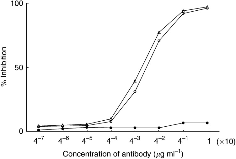
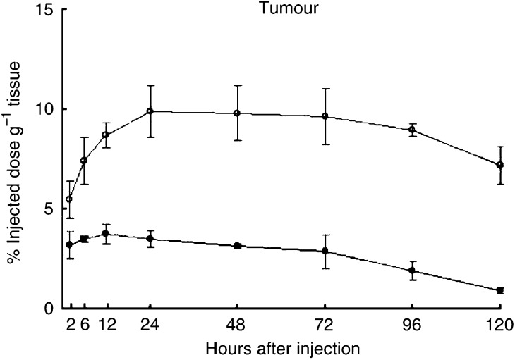
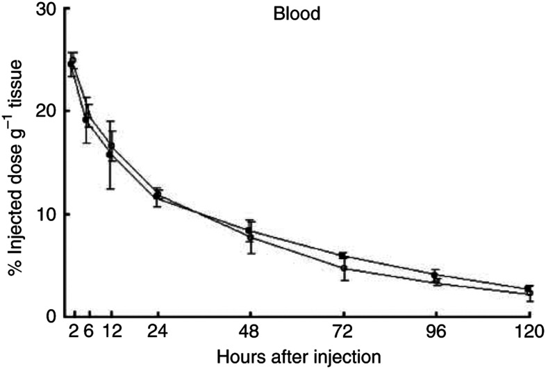
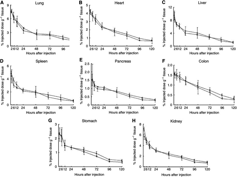
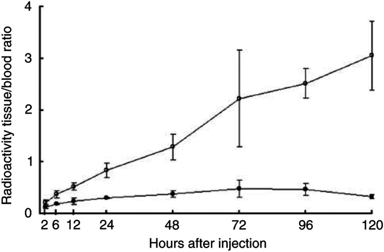
Superparamagnetic iron oxide (SPIO)-based colloid has been used clinically as a tissue-specific magnetic resonance contrast agent. We coupled monoclonal antibody A7 (Mab A7), which reacts specifically with human colorectal carcinoma, to Ferumoxides (SPIO) and examined the accumulation of this conjugate in xenografted tumours in nude mice. We examined in vitro immunoreactivity of Mab A7 coupled to Ferumoxides and its in vivo distribution in nude mice with human colorectal carcinoma. Magnetic resonance imaging of tumour-bearing nude mice was performed 72 h after injection of A7-Ferumoxides. A7-Ferumoxides retained binding activities that were nearly identical to intact Mab A7. More of the radiolabelled A7-Ferumoxides accumulated in the tumour than normal mouse IgG-Ferumoxides from 12 h onwards after injection (P<0.05). Both A7-Ferumoxides and normal mouse IgG-Ferumoxides disappeared from blood linearly over time. The accumulation levels in normal tissue decreased linearly over time but were lower than levels in tumours from 6 h. In magnetic resonance T2-weighted imaging of the tumour-bearing nude mice, signal intensity was reduced at the margin of the tumour by injection of A7-Ferumoxides. Mab A7 coupled to Ferumoxides is potentially suitable as a magnetic resonance contrast agent for detecting local recurrence of rectal carcinoma.
基于超顺磁性氧化铁(SPIO)的胶体已在临床上用作组织特异性磁共振造影剂。我们将与人结肠直肠癌特异性反应的单克隆抗体A7(Mab A7)与菲立磁(SPIO)偶联,并检测了该缀合物在裸鼠异种移植肿瘤中的蓄积情况。我们检测了与菲立磁偶联的Mab A7的体外免疫反应性及其在患有人类结肠直肠癌的裸鼠体内的分布。在注射A7-菲立磁72小时后,对荷瘤裸鼠进行磁共振成像。A7-菲立磁保留了与完整Mab A7几乎相同的结合活性。注射后12小时起,肿瘤中积聚的放射性标记A7-菲立磁比正常小鼠IgG-菲立磁更多(P<0.05)。A7-菲立磁和正常小鼠IgG-菲立磁在血液中的含量均随时间呈线性下降。正常组织中的蓄积水平随时间呈线性下降,但从6小时起低于肿瘤中的水平。在荷瘤裸鼠的磁共振T2加权成像中,注射A7-菲立磁后肿瘤边缘的信号强度降低。与菲立磁偶联的Mab A7有可能适合作为检测直肠癌局部复发的磁共振造影剂。