Kim Ji Eun, Kim Beom Joon, Jeong Mi Sook, Seo Seong Jun, Kim Myeung Nam, Hong Chang Kwun, Ro Byung In
Department of Dermatology, College of Medicine, Chung Ang University, Korea.
J Korean Med Sci. 2005 Aug;20(4):649-54. doi: 10.3346/jkms.2005.20.4.649.
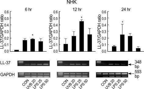
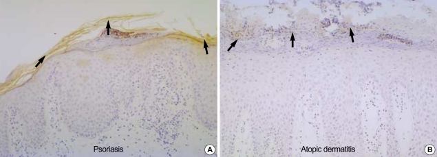
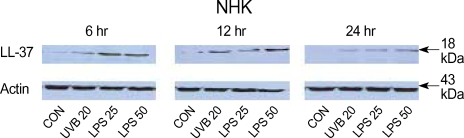
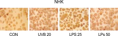
Defensins and cathelicidins (LL-37) are major antimicrobial peptides (AMPs) of the innate immune system of the human skin. In normal non-inflamed skin these peptides are negligible, but their expression can be markedly increased in inflammatory skin disease such as psoriasis. We designed this study to identify the expressions of LL-37 in normal human keratinocyte (NHK) and HaCaT cells after exposure to stimulants and to investigate difference of LL-37 expression accompanied with cell differentiation status, and come to understand difference of susceptibility to infection in atopic dermatitis and psoriasis. Expressions of LL-37 in NHKs and HaCaT cells were evaluated by using RT-PCR, Western blotting, and immunohistochemical (IHC) staining at 6, 12, and 24 hr post stimulation after exposure to Ultraviolet B irradiation and lipopolysaccharide. And expression of LL-37 in skin biopsy specimens from patients with atopic dermatitis and psoriasis was determined by immunohistochemical analysis. In time-sequential analyses of LL-37 expression revealed that LL-37 was expressed in NHKs, but not in HaCaT cells. IHC analysis confirmed the presence of abundant LL-37 in the epidermis of psoriasis. Therefore we deduced that expression of LL-37 is affected by UV irradiation, bacterial infection, and status of cell differentiation.
防御素和杀菌肽(LL-37)是人类皮肤固有免疫系统的主要抗菌肽。在正常非炎症性皮肤中,这些肽的含量可忽略不计,但在诸如银屑病等炎症性皮肤病中其表达会显著增加。我们设计本研究以确定正常人角质形成细胞(NHK)和HaCaT细胞在暴露于刺激物后LL-37的表达情况,并研究LL-37表达随细胞分化状态的差异,进而了解特应性皮炎和银屑病对感染易感性的差异。在暴露于紫外线B照射和脂多糖后,于刺激后6小时、12小时和24小时,通过逆转录聚合酶链反应(RT-PCR)、蛋白质免疫印迹法和免疫组织化学(IHC)染色评估NHK细胞和HaCaT细胞中LL-37的表达。并通过免疫组织化学分析确定特应性皮炎和银屑病患者皮肤活检标本中LL-37的表达。对LL-37表达的时序分析显示,LL-37在NHK细胞中表达,但在HaCaT细胞中不表达。免疫组织化学分析证实银屑病表皮中存在大量LL-37。因此我们推断LL-37的表达受紫外线照射、细菌感染和细胞分化状态的影响。