Burbelo Peter D, Goldman Radoslav, Mattson Thomas L
Lombardi Comprehensive Cancer Center, Department of Oncology, Georgetown University Medical Center, Washington, DC 20057, USA.
BMC Biotechnol. 2005 Aug 18;5:22. doi: 10.1186/1472-6750-5-22.
Assays detecting human antigen-specific antibodies are medically useful. However, the usefulness of existing simple immunoassay formats is limited by technical considerations such as sera antibodies to contaminants in insufficiently pure antigen, a problem likely exacerbated when antigen panels are screened to obtain clinically useful data.
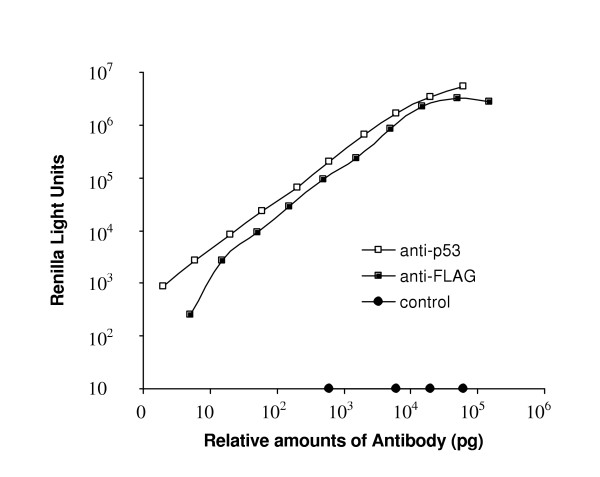
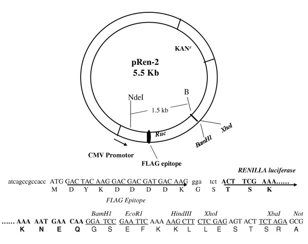
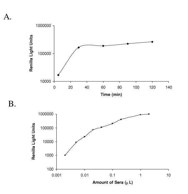
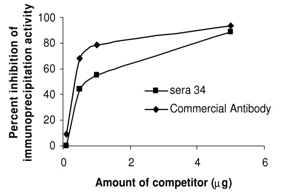
We developed a novel and simple immunoprecipitation technology for identifying clinical sera containing antigen-specific antibodies and for generating quantitative antibody response profiles. This method is based on fusing protein antigens to an enzyme reporter, Renilla luciferase (Ruc), and expressing these fusions in mammalian cells, where mammalian-specific post-translational modifications can be added. After mixing crude extracts, sera and protein A/G beads together and incubating, during which the Ruc-antigen fusion become immobilized on the A/G beads, antigen-specific antibody is quantitated by washing the beads and adding coelenterazine substrate and measuring light production. We have characterized this technology with sera from patients having three different types of cancers. We show that 20-85% of these sera contain significant titers of antibodies against at least one of five frequently mutated and/or overexpressed tumor-associated proteins. Five of six colon cancer sera tested gave responses that were statistically significantly greater than the average plus three standard deviations of 10 control sera. The results of competition experiments, preincubating positive sera with unmodified E. coli-produced antigens, varied dramatically.
This technology has several advantages over current quantitative immunoassays including its relative simplicity, its avoidance of problems associated with E. coli-produced antigens and its use of antigens that can carry mammalian or disease-specific post-translational modifications. This assay should be generally useful for analyzing sera for antibodies recognizing any protein or its post-translational modifications.
检测人类抗原特异性抗体的检测方法具有医学实用性。然而,现有的简单免疫分析方法的实用性受到技术因素的限制,例如血清抗体对纯度不足的抗原中的污染物的反应,当筛选抗原库以获得临床有用数据时,这个问题可能会更加严重。
我们开发了一种新颖且简单的免疫沉淀技术,用于鉴定含有抗原特异性抗体的临床血清,并生成定量抗体反应谱。该方法基于将蛋白质抗原与一种酶报告基因——海肾荧光素酶(Ruc)融合,并在哺乳动物细胞中表达这些融合蛋白,在该细胞中可以添加哺乳动物特异性的翻译后修饰。将粗提物、血清和蛋白A/G珠混合在一起并孵育,在此过程中Ruc-抗原融合蛋白固定在A/G珠上,通过洗涤珠子、添加腔肠素底物并测量发光来定量抗原特异性抗体。我们用患有三种不同类型癌症的患者的血清对这项技术进行了表征。我们发现,这些血清中有20%-85%含有针对五种频繁突变和/或过表达的肿瘤相关蛋白中至少一种的高滴度抗体。所检测的六份结肠癌血清中有五份的反应在统计学上显著高于10份对照血清的平均值加三个标准差。用未修饰的大肠杆菌产生的抗原预孵育阳性血清的竞争实验结果差异很大。
与目前的定量免疫分析方法相比,这项技术有几个优点,包括相对简单、避免了与大肠杆菌产生的抗原相关的问题以及使用了可以携带哺乳动物或疾病特异性翻译后修饰的抗原。这种检测方法通常应有助于分析血清中识别任何蛋白质或其翻译后修饰的抗体。